BIOLOGY Volume 1 - A Guide to General Biology - 2004
9. UTILIZATION OF ENERGY
9.3. Cell Respiration
9.3.10. Mitochondria
Cell/35.html">Mitochondria are present in all Eukaryotic Cells. These Organelles are the principal site of The Cell's aerobic respiratory activity. Mitochondria were first observed as granules in Muscle cells in 1850.
The number of mitochondria in a cell varies considerably, depending on the Organism and the type of cell. Cells with high energy demands contain numerous mitochondria (a rat Liver cell, for instance, may contain about 1,000). In less active cells, mitochondria are much fewer in number. The size and shape of mitochondria also vary widely. Mitochondria may be spiral, spherical, elongated, cup-shaped, or even branched; in more active cells, they are typically larger. The length of mitochondria ranges from 1.5 to 10 µm, and their width from 0.25 to 1.00 µm, but their diameter does not exceed 1 µm.
9.6. Why is the diameter of mitochondria relatively constant despite such large variations in their length?
Mitochondria are capable of changing shape, and some can also move to particularly active Regions of the cell. Such movement allows the cell to concentrate A large number of mitochondria in areas where the demand for ATP is highest. In other cases, THE POSITION OF mitochondria is more fixed (as, for example, in insect flight Muscles; Fig. 9.11).
Class="center">
Fig. 9.11. Transmission (A) and scanning (B) Cytology/cytology/93.html">ELECTRON MICROGRAPHS OF housefly flight muscle. Each myofibril (muscle fiber) can be seen surrounded by polymorphic (i.e., varying in shape) mitochondria. Myofibrils are the contractile elements of the muscle. Muscle contraction is an energy-requiring process.
Structure of Mitochondria
Mitochondria can be isolated from cells as a pure fraction using a homogenizer and ultracentrifuge, as described in Chapter 5 (Section 5.7). They can then be examined under an Electron microscope using various techniques, such as sectioning or negative staining.
Each mitochondrion is bounded by an envelope consisting of two membranes. The outer membrane is separated from the inner membrane by a small gap, the intermembrane space. The inner membrane forms numerous shelf-like folds known as cristae (Fig. 9.12). The cristae substantially increase the surface area of the inner membrane, providing a platform for the Components of the Respiratory Chain. The Active Transport of ADP and ATP takes place across The inner mitochondrial membrane. The negative staining technique, in which the surrounding space rather than the structures themselves is stained (Fig. 9.12, D), revealed the presence of distinct 'elementary particles' on the matrix-facing side of the inner mitochondrial membrane. Each such particle consists of a HEAD, a stalk, and a base (Fig. 9.12, C and D). Although micrographs (Fig. 9.12, E) might suggest that the elementary particles project from the membrane into the matrix, this is believed to be an artifact caused by the Sample preparation Procedure, and in reality they are fully embedded within the membrane. The heads of the particles are responsible for ATP synthesis; they contain the enzyme ATPase, which couples ADP phosphorylation to the Reactions of the respiratory chain. The components of the respiratory chain itself are located at the Base of the particles, spanning the entire thickness of the membrane. The mitochondrial matrix contains most of the Enzymes involved in the Krebs cycle, as well as those responsible for Fatty acid oxidation. It also houses Mitochondrial DNA, RNA, and 70S Ribosomes.


Fig. 9.12. Structure of mitochondria. A. Schematic diagram of a mitochondrion. B. Three-dimensional model. C. Diagram of crista structure with inner membrane 'elementary particles'. D. Structure of an elementary particle of the inner mitochondrial membrane. E. Low-magnification electron micrograph of a mitochondrion. F. High-magnification electron micrograph of a mitochondrion. G. Electron micrograph (transmission electron microscope; negative stain) showing elementary particles of the inner mitochondrial membrane from housefly mitochondria ruptured by osmotic Shock.
9.7. What chemical substances involved in Respiration are exchanged between the Cytoplasm and mitochondria? List four such substances entering the mitochondria and four substances passing from the mitochondria into the cytoplasm.
Evolution of Mitochondria: The Endosymbiotic Hypothesis
Like Bacteria (prokaryotes), mitochondria contain circular DNA (Fig. 9.13) and 70S ribosomes. This circumstance, along with other evidence, suggests that mitochondria, like METABOLISM/14.html">Chloroplasts, were once free-living bacteria. Having once accidentally invaded a Introduction/5.html">Eukaryotic Cell, they established a mutually beneficial Symbiosis with it. (We have discussed this topic in more detail previously; see Section 2.6.1.)
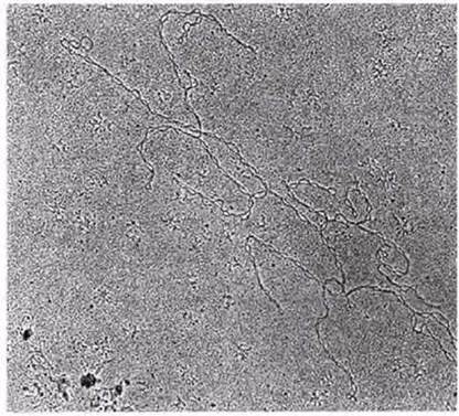
Fig. 9.13. Electron micrograph of mitochondrial DNA from the brewer's Yeast Saccharomyces carlsbergensis. The molecule is a supercoiled DNA ring with a circumference of 26 µm. It is composed of approximately 75,000 NUCLEOTIDES. It encodes certain mitochondrial Proteins. Other genes required by the mitochondria are located in the nuclear DNA.
Last update: 06/08/2026
Editorial and Educational Adaptation: This material has been compiled based on the primary/original source text. The project team performed an editorial review, corrected technical inaccuracies, structured sections, and adapted the content for an educational format.
What was processed:
- elimination of formatting defects (OCR errors, structural breaks, corrupted characters);
- editorial organization of content;
- standardization of terminology in accordance with academic sources;
- verification of factual statements against the original source text.
All mentions of the author, publication year, and origin of the primary text have been preserved in accordance with the source.