BIOLOGY Volume 1 - A Guide to General Biology - 2004
6. HISTOLOGY
6.3. Animal Epithelial Tissue
Animal Epithelial Tissue forms single-layer or multi-layered sheets that cover the internal and external surfaces of any Organism.
Epithelial Cells are connected to each other by a small amount of cementing substance, consisting mainly of CARBOHYDRATES, and specialized junctions known as intercellular contacts. The epithelium is underpinned by a basement membrane made of interwoven Collagen fibers embedded in a matrix. The term membrane should not be confused with The Cell membranes discussed in Chapter 5; here it simply denotes a thin layer. The matrix does not impede diffusion. Since epithelial cells lack a direct Blood supply, oxygen and nutrients reach them via diffusion from Lymphatic vessels located in the intercellular spaces. Nerve endings may also penetrate the epithelium.
The function of epithelial tissue is to protect underlying structures from mechanical damage and infection. Under constant mechanical stress, this tissue thickens and keratinizes; in areas where cells slough off due to persistent pressure or friction, Cell Division occurs at a very high rate, ensuring that lost cells are rapidly replaced. The free surface of the epithelium is often highly differentiated, performing absorptive, secretory, or excretory Functions, or housing sensory cells and nerve endings specialized for detecting stimuli.
Epithelial tissue is classified into several types (Table 6.2) depending on the number of cell layers and the shape of individual cells. In many PARTS OF THE body, cells of different types are intermingled, making it difficult to assign the epithelial tissue to any single definite category.
Class="center">Table 6.2. Types of Epithelial Tissue

6.3.1. Simple Epithelia
Squamous Epithelium
Squamous epithelial cells are thin and flattened—so much so that The Nucleus creates a bulge (Fig. 6.14). The cell edges are irregular, as clearly visible in surface views of the cell. Neighboring cells are tightly bound together by specialized junctions. Squamous epithelium is found in Bowman's capsules of the Kidneys, the lining of lung alveoli, and the walls of capillaries, where its thinness facilitates the diffusion of various substances. It also lines hollow structures such as Blood Vessels and Heart chambers, reducing friction as fluids flow through.


Fig. 6.14. Simple squamous epithelium. A. Schematic diagram. B. Light micrograph (small blood vessel). C. Transmission electron micrograph.
Cuboidal Epithelium
This is the least specialized of all epithelia. Its cells are cube-shaped and contain a centrally positioned spherical nucleus (Fig. 6.15). When viewed from above, the cells appear pentagonal or hexagonal in outline. Cuboidal epithelium lines the ducts of many glands, such as the Salivary Glands and Pancreas, as well as the proximal and distal convoluted tubules and collecting ducts of the Kidney in non-secretory regions.
Cuboidal epithelium is also present in many glands—salivary, mucous, sweat, and thyroid—where it performs secretory functions.

Fig. 6.15. Cuboidal epithelium. A. Schematic diagram. B. Light micrograph (kidney). C. Transmission electron micrograph.
Columnar Epithelium
The cells of this epithelium are tall and relatively narrow, a shape that provides a greater volume of Cytoplasm per unit surface area (Fig. 6.16). Each cell contains a nucleus located near its basal end. Goblet cells are frequently scattered among the epithelial cells; functionally, columnar epithelium can be secretory and/or absorptive. The free surface of each cell often features a well-defined brush border formed by microvilli, which increase the absorptive and secretory surface area. Columnar epithelium lines The Stomach, where mucus secreted by goblet cells protects the gastric mucosa from acidic contents and enzymatic Digestion. It also lines the intestine, where mucus likewise protects the intestinal walls from self-digestion while lubricating the passage of food. In the Small Intestine, digested nutrients are absorbed through this epithelium into the bloodstream. Columnar epithelium also lines and protects many renal tubules and is found in The Thyroid Gland and gall bladder.

Fig. 6.16. Columnar epithelium. A. Schematic diagram. B. Goblet cell. C. Light micrograph of the ileum epithelium showing a goblet cell. D. Transmission electron micrograph (ileum).
Ciliated Epithelium
The cells of this epithelium are typically columnar in shape, bearing numerous cilia on their free surfaces (Fig. 6.17). They are consistently associated with goblet cells that secrete mucus, which is propelled by ciliary beating. Ciliated epithelium lines the interior of the oviducts, Brain ventricles, central canal of the Spinal Cord, and respiratory tract (Trachea, Bronchi, and bronchioles), facilitating The transport of various substances. For instance, in the respiratory tract, the cilia sweep mucus upward toward the throat, which AIDS in clearing trapped particles. The mucus traps Bacteria, dust, and other fine particles, preventing them from entering the Lungs.
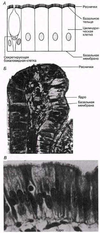
Fig. 6.17. Ciliated columnar epithelium. A. Schematic diagram. B. Photomicrograph of columnar epithelium lining the oviduct. C. Photomicrograph of columnar epithelium lining the trachea.
Pseudostratified epithelium
When examining histological sections of this epithelium, it appears that the cell nuclei lie at different levels because not all cells reach the free surface of the tissue (Fig. 6.18). Nevertheless, this epithelium consists of just a single layer of cells, each of which is attached to the basement membrane. Pseudostratified epithelium lines the Urinary Tract and the respiratory tract (trachea, bronchi, and bronchioles, where it is ciliated and composed of columnar cells).

Fig. 6.18. Pseudostratified epithelium. A. Columnar. B. Ciliated. C. Photomicrograph of ciliated epithelium of the respiratory tract. D. Scanning electron micrograph of cilia.
Last update: 06/08/2026
Editorial and Educational Adaptation: This material has been compiled based on the primary/original source text. The project team performed an editorial review, corrected technical inaccuracies, structured sections, and adapted the content for an educational format.
What was processed:
- elimination of formatting defects (OCR errors, structural breaks, corrupted characters);
- editorial organization of content;
- standardization of terminology in accordance with academic sources;
- verification of factual statements against the original source text.
All mentions of the author, publication year, and origin of the primary text have been preserved in accordance with the source.