BIOLOGY Volume 1 - A Guide to General Biology - 2004
6. HISTOLOGY
6.2. Plant Tissues Composed of Several Cell Types
6.2.2. Phloem
Phloem is similar to xylem in that it also contains tubular structures modified to carry out a transport function. However, these tubes are composed of living Cells containing Cytoplasm and do not serve a mechanical support function. Five distinct Cell types are recognized in the phloem: sieve elements (sieve tube members), companion cells, parenchyma cells, fibers, and sclereids.
Sieve Tubes and Companion Cells
Sieve tubes are long, tubular structures through which organic solutes—primarily sucrose solutions—are transported throughout the plant. They are formed by end-to-end alignment of cells known as sieve tube elements. In the apical meristem, where primary phloem and primary xylem (vascular bundles) originate, The Development of these cell rows from procambial strands can readily be observed.
The first-formed phloem, termed protophloem, appears—much like protoxylem—in the growing and elongating region of the ROOT or stem. As the surrounding Tissues expand, the protophloem is stretched, and a significant portion of it eventually dies off, ceasing to function. Simultaneously, however, new phloem is generated. This tissue, which matures after elongation has ceased, is called metaphloem.
Sieve tube elements possess a highly distinctive Structure. They have thin cell walls composed of Cellulose and pectic substances, making them resemble parenchyma cells; however, their nuclei disintegrate upon maturation, and their cytoplasm is reduced to a thin peripheral layer pressed against The Cell wall. Despite lacking a Nucleus, sieve tube elements remain alive, though their survival depends entirely on the adjacent companion cells, with which they share a common meristematic mother cell. A sieve tube element and its companion cell function as a single integrated unit; the companion cell features dense, highly active cytoplasm. The ultrastructure of these cells, as revealed by Electron Microscopy, is detailed in Chapter 13.
A defining characteristic of sieve tubes is the presence of sieve plates, a feature immediately apparent under a Light Microscope. A sieve plate forms at the junction between the end walls of two adjacent sieve tube elements. Initially, plasmodesmata traverse these cell walls, but their channels subsequently widen into pores, transforming the end walls into sieve-like structures through which solutions flow from one element to the next. Sieve plates are distributed at regular intervals along the sieve tube, corresponding to individual elements. The ultrastructure of sieve tubes, companion cells, and phloem parenchyma, as revealed by electron microscopy, is illustrated in Fig. 6.13.
Secondary phloem, which develops (like secondary xylem) from the vascular cambium, closely resembles primary phloem in structure, differing mainly in the presence of strands of lignified fibers and parenchymatous medullary rays (Chapter 22). However, secondary phloem is neither as extensively developed as secondary xylem nor permanent, being subject to continuous renewal (Chapter 22).
Class="center">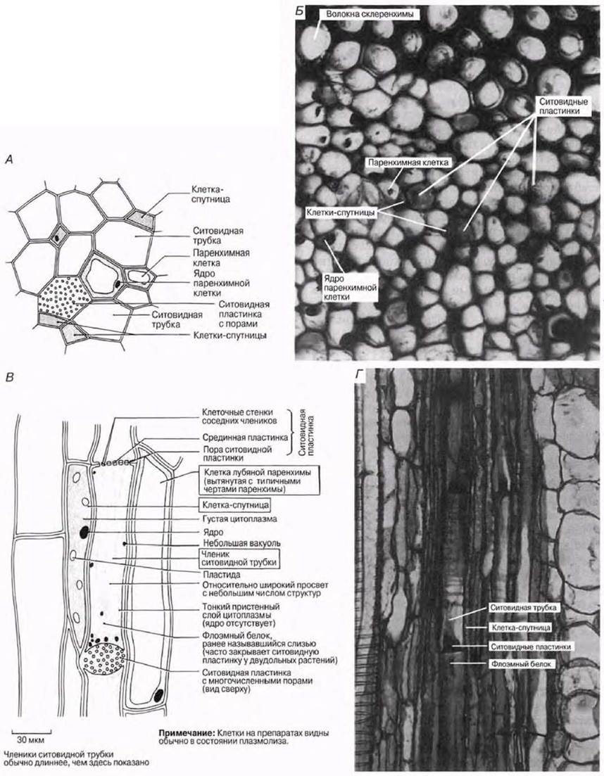
Fig. 6.13. STRUCTURE OF THE phloem. A. Schematic diagram of the phloem in transverse section. B. Photomicrograph of primary phloem in a stem cross-section of Helianthus; ×450. C. Schematic diagram of the phloem in longitudinal section. D. Photomicrograph of primary phloem in a stem Cytology/practical/54.html">Longitudinal section of Cucurbita; ×432.
Phloem Parenchyma, Phloem Fibers, and Sclereids
Phloem parenchyma and phloem fibers are present exclusively in dicotyledons and are absent in monocotyledons. Structurally, phloem parenchyma resembles Other types of parenchyma, though its cells are typically elongated. In secondary phloem, parenchyma occurs in the form of medullary rays and vertical files, much like the xylem parenchyma described above. Phloem and xylem parenchyma share identical Functions.
Phloem fibers are structurally indistinguishable from the sclerenchyma fibers described previously. While occasionally found in primary phloem, they are most commonly encountered in the secondary phloem of dicotyledons, where these cells form vertical strands. Because secondary phloem undergoes stretching during growth, sclerenchyma likely helps the tissue withstand these mechanical stresses.
Sclereids are frequently found within the phloem, particularly in older tissues.
Last update: 06/08/2026
Editorial and Educational Adaptation: This material has been compiled based on the primary/original source text. The project team performed an editorial review, corrected technical inaccuracies, structured sections, and adapted the content for an educational format.
What was processed:
- elimination of formatting defects (OCR errors, structural breaks, corrupted characters);
- editorial organization of content;
- standardization of terminology in accordance with academic sources;
- verification of factual statements against the original source text.
All mentions of the author, publication year, and origin of the primary text have been preserved in accordance with the source.