Human Biochemistry, Volume 2 - Murray R. 1993
Structure, Function, and Replication of Information Macromolecules
Protein Synthesis and the Genetic Code
Mutations
Mutations are alterations in The nucleotide sequence of a Gene. Even if a mutation initially occurs in the non-coding strand of a gene, one of the daughter molecules formed during Replication will necessarily contain the mutation at the corresponding position in the coding strand and give rise to a population of mutant Cells.
Base substitution mutations
Two Types of base substitutions are distinguished: transitions and transversions. Transitions refer to the replacement of a purine base by another purine, or a pyrimidine by another pyrimidine. Transversions are defined as the replacement of a purine base by a pyrimidine, or a pyrimidine base by a purine (Fig. 40.3).
If the nucleotide sequence of a gene containing a substitution is transcribed, the resulting mRNA molecule will have a complementary Substitution at the corresponding locus. A point substitution in an mRNA molecule during Translation into an Amino Acid Sequence can lead to various consequences:
1) if the substitution occurs at the third nucleotide of a codon, due to the degeneracy of METABOLISM/28.html">The Genetic Code, There is a probability that The amino acid sequence will remain unchanged and the mutation will not manifest itself in any way;
2) a missense effect may occur, where one amino acid is replaced by another As a result of a nucleotide substitution. Depending on its localization within the amino acid sequence of the protein, such a substitution may be acceptable, partially acceptable, or unacceptable with respect to the function of the given protein. An Analysis of the genetic code suggests that point mutations will most frequently lead to replacements by Amino Acids with fairly similar functional groups. If an acceptable substitution occurs, the protein molecule may turn out to be functionally indistinguishable from the normal one. As a result of a partially acceptable substitution, the normal functioning of the protein is impaired. Finally, an unacceptable substitution leads to a complete loss of its function;
Class="center">
Fig. 40.3. Scheme of the occurrence of transitions and transversions.
3) as a result of a point mutation, a nonsense codon may arise, the presence of which leads to premature termination of Protein Synthesis. As a rule, the fragment formed as a result of premature termination is incapable of performing the function of the intact protein molecule.
Mutations of globin genes
The Role of mutations can be conveniently analyzed using Hemoglobin genes as an example. A large body of factual material regarding the Amino acid sequences of normal and altered Hemoglobins has been accumulated in this field (see ch. 6). The hemoglobin molecule serves as a good example to demonstrate The Effect of point Amino Acid Substitutions and to illustrate some of the Selection/11.html">General features of the genetic code discussed earlier.
Some mutations are not overtly manifested. The lack of effect of individual point mutations can be directly demonstrated only by determining the nucleotide sequence of the hemoglobin gene or the corresponding mRNAs in A large number of individuals with normal hemoglobin. However, based on indirect data, it can be inferred that in the gene for the ß-chain, codon 67 (which encodes valine) is not identical in all individuals with normal ß-globin. In Milwaukee-type hemoglobin, glutamic acid is located at position 67 of the ß-chain, whereas in Bristol hemoglobin, it is aspartic acid. If the substitutions at position 67 of the ß-globin amino acid sequence are considered to be consequences of single-nucleotide substitutions in the corresponding mRNA codon, the aspartic acid codons GAU or GAC of Bristol hemoglobin could have been preceded prior to the point mutation by the valine codons GUU or GUC. At the same time, the glutamic acid codons GAA or GAG in the mRNA of Milwaukee hemoglobin must have been preceded by the valine codons GUA or GUG. Sydney-type hemoglobin, which has an Alanine at position 67 (codons GCU, GCC, GCA, or GCG), could have arisen as a result of a single-nucleotide substitution of any of the four valine codons (GUU, GUC, GUA, or GUG) (Fig. 40.4).
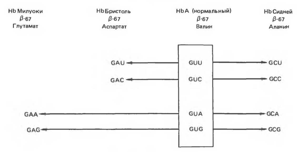
Fig. 40.4. In the normal ß-chain of human hemoglobin A, position 67 is occupied by valine encoded by one of four codons enclosed in the box. In abnormal Milwaukee-type hemoglobin, glutamate is found at this position, encoded by either the GAA or GAG codon. Both codons can arise as a result of a single transversion of the valine codons GUA or GUG. Similarly, alanine at position 67 of the ß-chain in Sydney-type hemoglobin can be the result of a single-nucleotide substitution in any of the four valine codons. Aspartic acid in Bristol-type hemoglobin can appear as a result of the substitution of a single nucleotide in one of the two valine codons, GUU or GUC.
Missense mutations
A. Acceptable missense mutations. An example of acceptable missense mutations in the structural gene of the hemoglobin ß-chain (Fig. 40.5, top) is a mutation detected by A change in the electrophoretic mobility of erythrocyte hemoglobin in practically healthy individuals. In members of at least two Japanese families, a hemoglobin variant called hemoglobin Hikari has been identified. In this type of hemoglobin molecule, asparagine replaces Lysine at position 61 of the ß-chain. The corresponding AAA or AAG codon is altered by a single-nucleotide transversion to AAU or AAC. The replacement of lysine by asparagine does not affect the normal function of the hemoglobin ß-chain in Hikari.

Fig. 40.5. Examples of Three types of missense mutations leading to the appearance of abnormal hemoglobin ß-chains. The amino acid substitutions and possible substitutions in the corresponding codons are indicated in the figure. The ß-chain of hemoglobin Hikari exhibits virtually normal physiological Functions with altered electrophoretic mobility. The function of hemoglobin S is partially impaired as a result of a mutation in the ß-chain: it can bind oxygen, but precipitates upon deoxygenation. In hemoglobin M Boston, as a result of a mutation in the a-chain, the Fe2+ ion incorporated into the heme is oxidized to Fe3+, which completely precludes oxygen binding.
B. Partially acceptable missense mutations. Partially acceptable missense mutations are best illustrated by sickle-Cell hemoglobin S (Fig. 40.5, middle). A missense mutation in the 6th codon of the hemoglobin ß-chain leads to the replacement of glutamic acid by valine (codons GUA or GUG are formed instead of GAA or GAG). Such a substitution interferes with the normal functioning of hemoglobin and, in the homozygous state, leads to Sickle-Cell Anemia. The replacement of glutamine by valine can be regarded as partially acceptable because the altered hemoglobin binds and releases oxygen, albeit abnormally.
C. Unacceptable missense mutations. Unacceptable missense mutations (Fig. 40.5, bottom) lead to The formation of completely non-functional hemoglobin. For example, a mutation in the hemoglobin M gene causes the Fe2+ ion within the heme to be oxidized to Fe3+, converting hemoglobin into the met-form. Methemoglobin is incapable of transporting oxygen (see ch. 6).
Frameshift mutations
This type of mutation is caused by deletions or insertions of NUCLEOTIDES into the gene sequence, which accordingly alters The sequence of the mRNA transcribed from it. The deletion of a single nucleotide in the coding strand leads to a frameshift in the mRNA. Since there are no codon-delimiting punctuation marks in the mRNA sequence, the translation machinery does not recognize the deletion. This results in the synthesis of a completely different polypeptide chain (Fig. 40.6, example 1), including in the region well distant from the deletion itself. As a result of a single-nucleotide deletion or insertion, the translated information is profoundly distorted; furthermore, nonsense codons may arise that terminate the further growth of the polypeptide chain (Fig. 40.6, example 3).
If three nucleotides, or a multiple of three, are deleted, protein molecules lacking a specific number of amino acids will be transcribed from the corresponding mRNA (Fig. 40.6, example 2). Because the genetic code is triplet-based, the amino acid sequence in this case will remain undistorted in the region distal to the deletion. If a deletion of one or two nucleotides occurs immediately before or within the stop codon, an abnormal elongation of the polypeptide chain may be observed. Translation will continue until the nearest stop codon (Fig. 40.6, example 1). Striking examples of such mutations are given in the Structure/133.html">Discussion of various hemoglobinopathies.
Insertions of one, two, or any number of nucleotides not a multiple of three into a gene also lead to the formation of an altered mRNA with a frameshift, which in turn leads to consequences fundamentally no different from those arising from deletions. These may include distortion of the amino acid sequence over an extended region downstream of the insertion site; the formation of a nonsense codon (at the insertion site or some distance from it) and premature termination of protein synthesis; or readthrough translation due to the elimination of the normal stop codon. An insertion occurring in the gene downstream of a deletion (or vice versa) can restore the correct reading frame (Fig. 40.6, example 4). Translation of such an mRNA will result in a polypeptide with a distorted segment enclosed between the insertion and deletion sites. Downstream of the reading frame restoration point, the amino acid sequence will be normal. One can envision numerous combinations of deletions and insertions that produce Proteins containing regions with altered structure flanked by regions with the original amino acid sequence. This phenomenon was convincingly demonstrated in bacteriophage T4, making a significant contribution to proving the triplet Nature of the genetic code.
Suppressor tRNA molecules
Above, we discussed the appearance of altered protein products resulting from structural Gene Mutations, assuming that all tRNA molecules function normally. However, abnormally functioning tRNAs, which are themselves the result of mutations, have been discovered in prokaryotes and lower eukaryotes. Some of these anomalous tRNA molecules are capable of suppressing structural gene mutations. Suppressor tRNA molecules typically arise from alterations within the anticodon region. They can suppress missense, nonsense, and frameshift mutations. However, because suppressor tRNAs cannot distinguish between a normal codon and a mutation-generated codon, their presence in The Cell is usually accompanied by reduced viability. For example, a nonsense suppressor tRNA will also suppress normal termination signals, thereby permitting undesirable readthrough of normal genes. Frameshift suppressor tRNA molecules can read a normal codon and the first nucleotide of the subsequent codon, leading to a reading frame shift even when undesirable. Suppressor tRNAs likely also exist in mammalian cells, as The phenomenon of stop-codon readthrough has been observed in them.
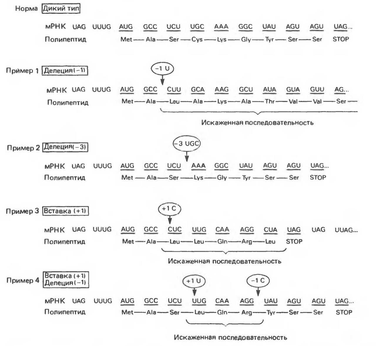
Fig. 40.6. Examples of various variant changes in mRNA structure and the translated amino acid sequence caused by deletions and insertions within the coding region of a gene. Arrows indicate insertion and deletion sites. Numbers in circles indicate the number of inserted or deleted nucleotides.
Last update: 06/08/2026
Editorial and Educational Adaptation: This material has been compiled based on the primary/original source text. The project team performed an editorial review, corrected technical inaccuracies, structured sections, and adapted the content for an educational format.
What was processed:
- elimination of formatting defects (OCR errors, structural breaks, corrupted characters);
- editorial organization of content;
- standardization of terminology in accordance with academic sources;
- verification of factual statements against the original source text.
All mentions of the author, publication year, and origin of the primary text have been preserved in accordance with the source.