Principles of Biochemistry Volume 1 - A. Lehninger 1985
Biomolecules
Lipids and Membranes
Erythrocyte membranes have been studied in great detail
The Study of Proteins contained in the erythrocyte Cell/33.html">Plasma Membrane has provided new insights into Membrane Structure, leading to the hypothesis that at least some membranes possess a «Skeleton». The human erythrocyte membrane contains five major proteins and A large number of minor ones. Most Membrane Proteins are Glycoproteins. Glycophorin (a «sugar carrier») is one of the integral proteins of The erythrocyte membrane. It has a Molecular Weight of 30,000 and consists of 130 amino acid residues alongside numerous sugar residues, which account for about 60% of the entire molecule. At one end of the polypeptide chain lies a complex hydrophilic HEAD comprising up to 15 oligosaccharide chains, each consisting of approximately 10 sugar residues. At the other end of the glycophorin polypeptide chain, There is a large number of glutamic and aspartic acid residues (Fig. 12-20) that carry a negative charge at pH 7.0. In the middle of the molecule, between the two hydrophilic ends, lies a segment of the polypeptide chain containing about 30 hydrophobic amino acid residues. The sugar-rich end of the glycophorin molecule is localized on the outer surface of the erythrocyte membrane, protruding from it like a small bush. It is believed that the hydrophobic segment located in the middle of the glycophorin molecule spans The Lipid Bilayer, while the polar end with its negatively charged amino acid residues is immersed in the Cytosol. The sugar-rich head of glycophorin contains antigenic determinants that determine Blood Groups (A, B, or O). In addition, it features binding sites for certain pathogenic Viruses.
Another major erythrocyte membrane protein, spectrin, accounts for up to 20% of the total membrane protein. This peripheral protein is located on the inner surface of the membrane and is readily extracted. The spectrin molecule consists of four polypeptide chains with a total molecular weight of approximately 1 million; these chains form long, flexible rods 100-200 nm in length. By binding to specific proteins and Lipids on the inner surface of the erythrocyte membrane, spectrin molecules form a flexible lattice that presumably acts as a membrane skeleton. Actin microfilaments also bind to spectrin, and it is highly likely that they link the spectrin rods to one another. Thus, it can be said that the erythrocyte membrane possesses a skeleton, or framework, to which specific lipids and membrane proteins are anchored (Fig. 12-21).
Class="center">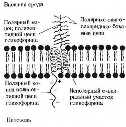
Fig. 12-20. The glycophorin molecule in the erythrocyte membrane. The branched carbohydrate chains protruding from the membrane bear specific sites that determine blood groups, as well as sites responsible for binding certain viruses.
The Plasma Membranes of other Cells have a more complex structure. On the outer surface of cells in many dense Tissues, there is another important glycoprotein, Fibronectin (Sec. 11.12), which exhibits high adhesive capacity and may mediate the adhesion of Cells of the same type to one another.
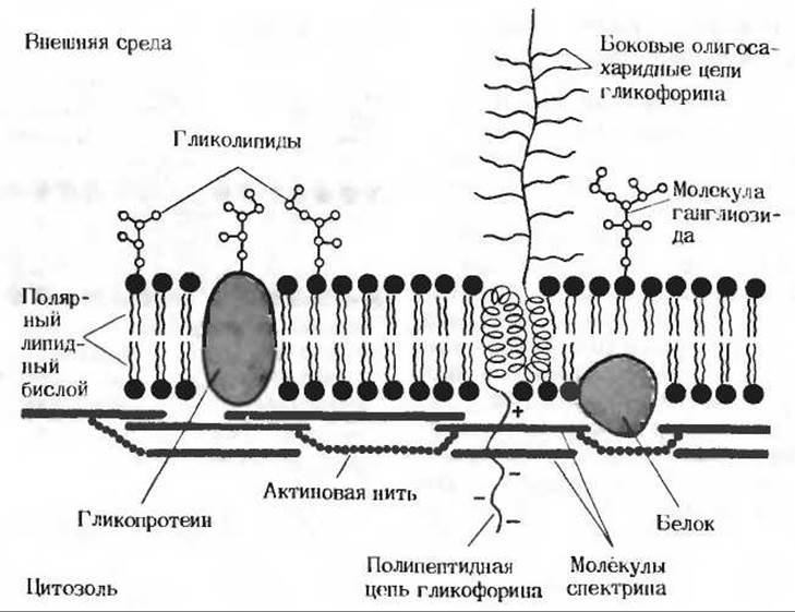
Fig. 12-21. Schematic representation of a section of the erythrocyte membrane. The diagram shows oligosaccharide «antennae» formed by membrane glycoproteins and Glycolipids, side oligosaccharide chains of glycophorin, and a skeletal framework attached to the inner surface of the membrane, consisting of spectrin molecules linked together by short actin filaments.
Last update: 06/08/2026
Editorial and Educational Adaptation: This material has been compiled based on the primary/original source text. The project team performed an editorial review, corrected technical inaccuracies, structured sections, and adapted the content for an educational format.
What was processed:
- elimination of formatting defects (OCR errors, structural breaks, corrupted characters);
- editorial organization of content;
- standardization of terminology in accordance with academic sources;
- verification of factual statements against the original source text.
All mentions of the author, publication year, and origin of the primary text have been preserved in accordance with the source.