Glycoproteins - Hughes R. 1985
Functions
Intracellular transport of glycoproteins
Lysosomal enzyme cycle
Data providing direct Evidence for the critical role of normal glycosylation has emerged over the past five years from studies on the intracellular lifecycle of a group of lysosomal Hydrolases. These investigations revealed a broadly significant system that also uncovers the biochemical defect underlying a specific human pathological condition.
This rare disorder is known as I-Cell disease. (Not to be confused with II Antigens; while this nomenclature is less than ideal, it is widely used in literature and not easily changed.) Individuals suffering from I-cell disease die at an early age, exhibiting massive intracellular accumulations of high-molecular-weight Glycoconjugates. The primary cause of this condition is a disruption in the normal levels of intracellular lysosomal hydrolases responsible for breaking down internalized Connective Tissue components. A similar defect is observed in cultured Cells (e.g., Skin fibroblasts) derived from such patients: these cultured cells secrete abnormally high amounts of lysosomal hydrolases into the medium. In other words, these cells secrete hydrolases that, under normal conditions, reside within lysosomal vesicles to degrade engulfed macromolecules and cellular debris [64]. Both normal and I-cells feature a surface receptor that recognizes specific Structure/135.html">Structural motifs on lysosomal hydrolases, mediating adsorptive endocytosis of these Enzymes. While I-cells can mediate the uptake of hydrolases from normal cells, they fail to recognize the enzymes synthesized and secreted by themselves. Moreover, normal cells are similarly unable to recognize hydrolases synthesized by I-cells; this clearly indicates that the observed defect relates to The structure of the I-cell hydrolases themselves, rather than an impairment in adsorptive endocytosis or cellular receptor function. The crucial role of the carbohydrate component in lysosomal hydrolases was first established upon discovering that 1) mannose-6-phosphate competitively inhibits the uptake of normal hydrolases by normal fibroblasts, 2) various lysosomal enzymes possess phosphorylated N-glycans, and 3) phosphatase Treatment leads to a reduction in hydrolase uptake. Enzymes secreted by I-cells lack this carbohydrate component, failing to bind to The surface of either normal or I-cells and remaining uninternalized. Mannose-6-phosphate receptors are present in low numbers on The Cell surface, which is where they were originally discovered. However, at least 90% of all receptors reside within The Endoplasmic reticulum, Golgi membranes, and Lysosomes. This suggests that The primary function of these receptors is likely The transport of newly synthesized hydrolases from the rough endoplasmic reticulum to smooth membranes and, ultimately, to lysosomes (Fig. 4.7).
Class="center">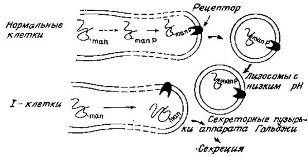
Fig. 4.7. Schematic diagram of Intracellular Transport of lysosomal hydrolases and the defect present in I-cells.
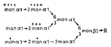
Fig. 4.8. Phosphorylated N-glycans of lysosomal enzymes [65]. The degree of phosphorylation of individual mannose residues is indicated by crosses.
The following sequence of events can be hypothesized. Following the glycosylation of newly synthesized Polypeptides with oligomannose intermediates, specific mannose residues undergo phosphorylation. This process is non-random, as certain mannose residues are preferentially phosphorylated (Fig. 4.8, +++), while others are not phosphorylated at all. In many cases, a single N-glycan may contain two phosphorylated mannose residues, and a given specific enzyme (e.g., ß-glucuronidase) may exhibit an entire array of glycan variants. The causes of this heterogeneity remain elusive [65]. Mannose-6-phosphate units are recognized by specific receptors localized on the cisternal face of those Endoplasmic reticulum and Golgi regions that ultimately bud off to form primary lysosomes, which contain a full Complement of hydrolytic enzymes on the inner surface of the lysosomal membrane. Lysosomes are characterized by a low pH, likely maintained by a high concentration of sialic acid-containing Glycoproteins projecting into the interior of the vesicles. This acidic pH weakens the interaction between mannose-6-phosphate groups and their specific receptors, allowing the hydrolases to become more or less freely distributed within the lysosomal fluid. Glycans at this stage are dephosphorylated to remove the recognition signal [66]. Macromolecules and cellular debris entering the cells via adsorptive endocytosis as endocytic vesicles subsequently fuse with primary lysosomes and are rapidly degraded by the complete battery of lysosomal enzymes (operating at an optimal pH of 4.5). In I-cells, the phosphorylation of N-glycans appears to be blocked. Consequently, recognition signals are absent, and lysosomal enzymes are misdirected into secretory vesicles rather than the endoplasmic reticulum and Golgi compartments destined for lysosome biogenesis.
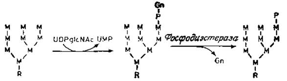
Fig. 4.9. Intermediates in The Biosynthesis of lysosomal ß-glucuronidase. R represents the core region of N-glycans. Only one phosphorylated form is shown; other forms are depicted in Fig. 4.8.
Recent experiments, however, have necessitated a revision of this simple model [67, 68]. The phosphorylation of mannose residues in lysosomal hydrolases does not occur directly via the Transfer of phosphate groups from ATP, such as through kinase action. Instead, following partial Processing and The transfer of a standard glucose- and mannose-containing polysaccharide from a lipid precursor to the protein (Fig. 4.9), an a-N-acetylglucosamine-1-phosphate moiety is transferred from UDP-N-acetylglucosamine to the C-6 position of a mannose residue. Because this reaction is highly selective and occurs exclusively with lysosomal enzymes, it presumably takes place only during a late stage of endoplasmic reticulum differentiation and lysosomal precursor formation. Furthermore, it is possible that only lysosomal enzymes possess a specific domain (a segment of the polypeptide chain) within their "recognition site" that excludes other high-mannose glycoproteins. It is also conceivable that the corresponding specific transferase is localized near mannose-6-phosphate receptors in specialized Regions of the endoplasmic reticulum on the cisternal side. In the subsequent enzymatic step, a highly specific a-N-acetylglucosaminyl phosphodiesterase generates the mannose-6-phosphate "signal," which releases free N-acetylglucosamine and triggers the binding of lysosomal enzymes to their receptors. Both described enzymes are predominantly found in purified smooth Golgi membrane preparations, which fits well with the overarching hypothesis. Consistent with the finding that the biochemical defect in I-cells stems from an inability to generate the phosphorylated form of lysosomal enzymes, the specific N-acetylglucosamine-1-phosphate transfer reaction is completely absent in these cells. As expected given that lysosomes are a characteristic cellular component, mannose-6-phosphate-containing enzymes are widely distributed.
Last update: 06/08/2026
Editorial and Educational Adaptation: This material has been compiled based on the primary/original source text. The project team performed an editorial review, corrected technical inaccuracies, structured sections, and adapted the content for an educational format.
What was processed:
- elimination of formatting defects (OCR errors, structural breaks, corrupted characters);
- editorial organization of content;
- standardization of terminology in accordance with academic sources;
- verification of factual statements against the original source text.
All mentions of the author, publication year, and origin of the primary text have been preserved in accordance with the source.