Biochemistry and Molecular Biology - Belyasova N.A. 2002
Structure and Functions of Cellular Components
Proteins. Organizational Features and Functions of Enzymes
Mechanism of Enzymatic Catalysis
The sequence of events in Enzymatic Catalysis can be described by the following scheme. First, an enzyme-substrate complex is formed. This process involves Conformational Changes in both the enzyme and substrate molecules, with the latter being held in the Active Site in a strained configuration. This forms an activated complex, or Transition State—a high-energy intermediate Structure that is less stable energetically than the initial reactants and products. A crucial contribution to the overall catalytic effect is made by transition-state stabilization, which involves interactions between the protein's amino acid residues and the strained substrate. The difference in Free energy between the initial reactants and the transition state corresponds to the activation energy (ΔG#). The reaction rate depends on the magnitude of ΔG#: the smaller it is, the higher the reaction rate, and vice versa. Essentially, ΔG# represents the "energy barrier" that must be overcome for the reaction to take place. Stabilization of the transition state lowers this "barrier" or activation energy. The actual chemical reaction occurs in the next step, after which the resulting products are released from the enzyme-product complex.
Several factors account for the high catalytic activity of Enzymes in lowering the reaction's energy barrier.
1. The enzyme can bind reacting substrate molecules in such a way that their reactive groups are positioned close to each other and to the catalytic groups of the enzyme (proximity effect).
2. The formation of the enzyme-substrate complex ensures substrate immobilization and optimal orientation for the Cleavage and formation of chemical bonds (orientation effect).
3. Substrate binding leads to the removal of its Hydration shell (which typically surrounds Water-soluble substances).
4. The induced-fit effect between the substrate and the enzyme.
5. Transition-state stabilization.
6. Specific groups within the enzyme molecule can provide Acid-Base Catalysis (proton transfer within the substrate) and Covalent Catalysis (formation of covalent bonds with the substrate, yielding more reactive structures than the substrate itself).
One classic example of acid-base catalysis is the Hydrolysis of glycosidic bonds in murein (peptidoglycan) by Lysozyme. Lysozyme is an enzyme found in the Cells of various animals and plants, including tears, saliva, egg white, and milk. Chicken egg white lysozyme has a Molecular Weight of 14,600 Da, consists of a single polypeptide chain (129 amino acid residues), and contains 4 disulfide bridges, which ensure its high stability. X-ray crystallographic Analysis of the lysozyme molecule has shown that it consists of two domains forming a "cleft" where the active site is located. A hexasaccharide binds along this cleft, with a specific subsites (A, B, C, D, E, and F) on the enzyme dedicated to binding each of the six sugar rings of murein (Fig. 6.4).
Class="center">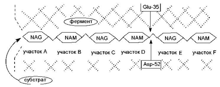
Fig. 6.4. Arrangement of the murein hexasaccharide unit in the Active Site of the lysozyme molecule: NAG stands for N-acetylglucosamine residue; NAM stands for N-acetylmuramic acid residue; the shaded area indicates the region of the lysozyme active site.
The murein molecule is held in the active site of lysozyme primarily through Hydrogen Bonds and hydrophobic interactions. Located in close proximity to the cleavage site of the glycosidic bond are two amino acid residues of the active site: glutamic acid at position 35 in the polypeptide chain, and aspartic acid at position 52 (Fig. 6.5).
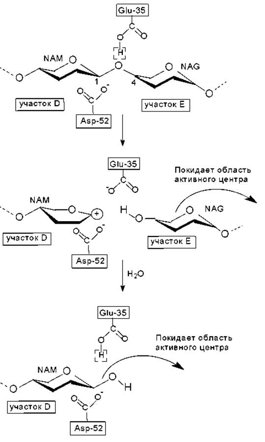
Fig. 6.5. Mechanism of glycosidic bond hydrolysis in the murein molecule facilitated by the enzyme lysozyme.
The side chains of these residues lie on opposite surfaces of the cleft, very close to the target glycosidic bond—at a distance of approximately 0.3 nm. The glutamate residue is situated in a nonpolar environment and is unprotonated, whereas the aspartate residue is in a polar environment, where its carboxyl group is deprotonated and acts as a hydrogen acceptor within an intricate network of hydrogen bonds.
The hydrolysis proceeds as follows. The protonated carboxyl group of Glu-35 donates its proton to the glycosidic oxygen atom, which leads to the Cleavage of the bond between this oxygen atom and the C1 atom of the sugar ring located in subsite D (general acid catalysis stage). This yields a product containing the sugar rings that were originally in
subsites E and F, which can then dissociate from the enzyme complex. The conformation of the sugar ring in subsite D is distorted into a half-chair conformation, in which five of the six atoms forming the ring lie virtually in the same plane. This structure corresponds to the transition-state conformation. Simultaneously, the C1 atom becomes positively charged, and this reaction intermediate is referred to as a carbonium ion (carbocation). The Free energy of the transition state is lowered through the stabilization of the carbocation by the deprotonated carboxyl group of Asp-52 (Fig. 6.5).
In the next step, a water molecule enters the reaction, replacing the disaccharide fragment that diffuses away from the active site. A proton from the water molecule is transferred to Glu-35, while a hydroxyl ion (OH-) attacks the C1 atom of the carbocation (general base catalysis stage). As a result, the second fragment of the cleaved polysaccharide becomes a reaction product (adopting a chair conformation) and leaves the active site, while the enzyme returns to its initial state, ready to carry out the next disaccharide cleavage reaction (Fig. 6.5).
Last update: 06/08/2026
Editorial and Educational Adaptation: This material has been compiled based on the primary/original source text. The project team performed an editorial review, corrected technical inaccuracies, structured sections, and adapted the content for an educational format.
What was processed:
- elimination of formatting defects (OCR errors, structural breaks, corrupted characters);
- editorial organization of content;
- standardization of terminology in accordance with academic sources;
- verification of factual statements against the original source text.
All mentions of the author, publication year, and origin of the primary text have been preserved in accordance with the source.