BIOCHEMISTRY: A TEXTBOOK FOR MEDICAL UNIVERSITIES - E.S. Severin - 2004
CHAPTER 7. CARBOHYDRATE METABOLISM
II. Carbohydrate Digestion
Intestinal epithelial Cells can absorb only Monosaccharides. Therefore, the digestive process consists of the Enzymatic Hydrolysis of glycosidic bonds in oligo- and polysaccharide CARBOHYDRATES (Fig. 7-10).
Class="center">Fig. 7-10. Hydrolysis of the glycosidic bond.

A. Carbohydrate Digestion IN THE Oral Cavity
In the oral cavity, food is mechanically broken down by chewing and mixed with saliva. Saliva is 99% Water and typically has a pH of 6.8. It contains the hydrolytic enzyme α-amylase (α-1,4-glucosidase), which cleaves α-1,4-glycosidic bonds in starch. Complete digestion of starch cannot occur in the oral cavity because the enzyme's action is brief. Furthermore, salivary amylase does not cleave α-1,6-glycosidic bonds (branch points); consequently, starch is only partially digested into large fragments called dextrins and a small amount of maltose. It should be noted that salivary amylase does not hydrolyze glycosidic bonds in Disaccharides.
The action of salivary amylase ceases in the strongly acidic environment of The Stomach contents (pH 1.5–2.5). However, within the food bolus, amylase activity may persist for some time until the pH drops. Gastric juice does not contain carbohydrate-digesting Enzymes. Only minor acid hydrolysis of glycosidic bonds may occur within the gastric contents.
B. Carbohydrate Digestion in the Intestine
Subsequent stages of digestion for undigested or partially digested starch, as well as other dietary carbohydrates, take place in various segments of the Small Intestine through the action of hydrolytic enzymes known as glycosidases.
Pancreatic α-Amylase
In the duodenum, the acidic pH of the stomach contents is neutralized because pancreatic secretion has a pH of 7.5–8.0 and contains bicarbonates (HCO3-). Pancreatic α-amylase enters the intestine via pancreatic secretions. This enzyme hydrolyzes α-1,4-glycosidic bonds in starch and dextrins.
The products of starch digestion at this stage include the disaccharide maltose, which consists of 2 glucose residues linked by an α-1,4 bond. Glucose residues located at the branch points of the starch molecule, joined by an α-1,6-glycosidic bond, form the disaccharide isomaltose. In addition, Oligosaccharides containing 3 to 8 glucose residues linked by α-1,4 and α-1,6 bonds are formed (Fig. 7-11).
Fig. 7-11. Hydrolysis of Starch by pancreatic α-amylase.

Pancreatic α-amylase, much like salivary amylase, acts as an endoglycosidase. Pancreatic α-amylase does not cleave α-1,6-glycosidic bonds in starch, nor does it hydrolyze the β-1,4-glycosidic bonds that link glucose residues in Cellulose. Consequently, cellulose passes through the intestine unchanged. Nevertheless, indigestible cellulose performs an important function as dietary fiber, adding bulk to food and positively influencing digestion. Additionally, in the Large Intestine, cellulose can be acted upon by bacterial enzymes and partially broken down to yield alcohols, organic acids, and CO2. These bacterial breakdown products are important stimulants of intestinal peristalsis.
Maltose, isomaltose, and trioses formed from starch in the upper intestine are intermediates. Their further digestion occurs through the action of specific enzymes in the small intestine. The dietary disaccharides sucrose and lactose are also hydrolyzed by specific disaccharidases in the small intestine.
A distinctive feature of carbohydrate digestion in the small intestine is that The activity of specific oligo- and disaccharidases in the intestinal lumen is low. Instead, these enzymes act actively On the surface of intestinal epithelial cells.
The inner lining of the small intestine features finger-like projections called villi, which are covered with epithelial cells. These epithelial cells, in turn, are covered with microvilli facing the intestinal lumen. Together with the villi, these cells form the brush border, which significantly increases the contact surface area between hydrolytic enzymes and their substrates in the intestinal contents. In humans, there are 80–140 million villi per 1 mm2 of the small intestine surface.
The enzymes that cleave glycosidic bonds in disaccharides (disaccharidases) form enzymatic complexes localized on the outer surface of the enterocyte cytoplasmic membrane.
Sucrase-Isomaltase Complex
This enzymatic complex consists of two polypeptide chains and features a domain Structure. The sucrase-isomaltase complex is anchored to the microvillar membrane of the intestine via a hydrophobic (transmembrane) domain formed by the N-terminal region of the polypeptide. The catalytic domain projects into the intestinal lumen (Fig. 7-12). This membrane attachment facilitates the efficient cellular uptake of hydrolysis products.
Fig. 7-12. Sucrase-isomaltase complex. 1 — sucrase; 2 — isomaltase; 3 — binding domain; 4 — transmembrane domain; 5 — cytoplasmic domain.

The sucrase-isomaltase complex hydrolyzes sucrose and isomaltose by cleaving α-1,2 and α-1,6-glycosidic bonds. In addition, both enzyme domains exhibit maltase and maltotase activities, hydrolyzing α-1,4-glycosidic bonds in maltose and maltotriose (a trisaccharide derived from starch). The sucrase-isomaltase complex accounts for 80% of the total intestinal maltase activity. Despite this high intrinsic maltase activity, the enzyme complex is named after its primary Specificity. Furthermore, the sucrase subunit is the sole intestinal enzyme capable of hydrolyzing sucrose. The isomaltase subunit hydrolyzes glycosidic bonds in isomaltose at a higher rate than in maltose and maltotriose (Figs. 7-13, 7-14).
Fig. 7-13. Action of the sucrase-isomaltase complex on maltose and maltotriose.

Fig. 7-14. Action of the sucrase-isomaltase complex on isomaltose and oligosaccharide.

The content of the sucrase-isomaltase enzyme complex is quite high in the jejunum, but decreases in the proximal and distal PARTS OF THE intestine.
Glucoamylase complex
This enzyme complex catalyzes the hydrolysis of α-1,4 linkages between glucose residues in oligosaccharides, acting from the reducing end. Based on its MECHANISM OF ACTION, the enzyme is classified as an exoglycosidase. The complex also cleaves bonds in maltose, functioning as a maltase. The glucoamylase complex comprises two distinct catalytic subunits with minor differences in substrate specificity. The glucoamylase activity of the complex is highest in the lower Regions of the small intestine.
β - Glycosidase complex (lactase)
Lactase cleaves the β-1,4-glycosidic bonds between galactose and glucose in lactose (Fig. 7-15).
Fig. 7-15. Action of lactase.

Chemically, this enzyme complex is a glycoprotein. Like other glycosidase complexes, lactase is associated with the brush border and is distributed unevenly throughout the small intestine. Lactase activity varies depending on age. For instance, fetal lactase activity is particularly elevated during late gestation and remains high until 5–7 years of age. Subsequently, the enzyme activity declines, dropping in adults to 10% of the activity level characteristic of children.
Trehalase is another glycosidase complex that hydrolyzes the bonds between monomers in trehalose, a disaccharide found in mushrooms. Trehalose consists of two glucose residues linked by a glycosidic bond between their first anomeric carbon atoms (Fig. 7-16).
Fig. 7-16. Structure of trehalose.

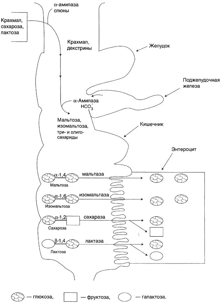
The concerted action of all these enzymes completes the Digestion of dietary oligo- and Polysaccharides, yielding monosaccharides, predominantly glucose. In addition to glucose, dietary carbohydrates also give rise to FRUCTOSE AND GALACTOSE, and in smaller amounts, mannose, xylose, and arabinose. The overall pathway of carbohydrate digestion is illustrated in Fig. 7-17.
Fig. 7-17. Digestion of carbohydrates.
Last update: 06/08/2026
Editorial and Educational Adaptation: This material has been compiled based on the primary/original source text. The project team performed an editorial review, corrected technical inaccuracies, structured sections, and adapted the content for an educational format.
What was processed:
- elimination of formatting defects (OCR errors, structural breaks, corrupted characters);
- editorial organization of content;
- standardization of terminology in accordance with academic sources;
- verification of factual statements against the original source text.
All mentions of the author, publication year, and origin of the primary text have been preserved in accordance with the source.