Amino Acids, Peptides and Proteins - Dévényi T., Gergely J. 1976
Methods of medium- and high-voltage electrophoresis
Apparatus for medium-voltage horizontal electrophoresis and its application
Electrophoresis is carried out at low Temperature in small volumes of buffer solution on horizontally positioned filter paper at a voltage gradient of 30—40 V/cm.
1. Description of the apparatus. Fig. 15 schematically illustrates a custom-built horizontal electrophoresis apparatus equipped with a 44x44 cm Plexiglas cooling plate. The Circulation of the coolant inside the plate is directed by special baffles. A 50% ethanol solution, cooled in a special reservoir with an ice-acetone mixture or by a refrigeration unit, is used as the coolant. The temperature of the cooling plate should be approximately +6° C. A sheet of filter paper moistened with buffer solution is placed on the plate and connected to the electrode compartments of the apparatus by cellophane-covered paper wicks. The filter paper sheet is covered from above with a 15 mm thick Plexiglas plate, which is tightly pressed against the cooling plate at three points.
The electrode compartment of the apparatus is a rectangular Plexiglas chamber divided into two sections. The outer section contains an electrode made of 0.3 mm thick platinum wire; the inner section accommodates the wicks connecting the compartment to the filter paper. Both sections of the electrode compartment are connected by a bridge of wet filter paper.
The power supply is equipped with a continuously variable rheostat and provides a maximum voltage of 2000 V and a current of 150—200 mA.
The coolant is circulated through the system by a small pump.
2. Preparation of Buffer solutions. 1) Buffer solution pH 4.3— 5.0 (the pH value is determined by the purity of the acetic acid): mix 10 ml of acetic acid and 10 ml of pyridine and dilute with distilled Water to 1000 ml.
2) Buffer solution pH 6.5: mix 100 ml of pyridine and 4 ml of acetic acid and dilute with distilled water to 1000 ml.
3) Buffer solution pH 2.0: mix 100 ml of acetic acid and 35 ml of formic acid and dilute with distilled water to 1000 ml.
Class="center">
Fig. 15. Schematic diagram of an apparatus for horizontal medium-voltage paper electrophoresis with cooling.
A. 1 — cooling plate (see B); 2 — plate serving as a cover; 3 — sheet of filter paper; 4 — cellophane membrane; 5 — filter paper wick; 6 — filter paper bridge; 7 — electrode compartment; 8 — cooling plate holders; 9 — fastening detail; 10 — sponge rubber strip; 11 — platinum electrodes.
3. Treatment of filter paper. The necessary markings are made on a sheet of filter paper, which is then moistened with the appropriate buffer solution. After carefully blotting the excess buffer solution between several layers of filter paper, the sheet is placed on the cooling plate. Then, covered with a dry sheet of paper, it is smoothed out with a rubber roller of the type used in photographic laboratories. Cellophane strips, slightly longer than the width of the paper, are placed on the ends of the filter paper sheet facing the electrodes. The cellophane strip is folded so that it wraps around the top and bottom of the end of the paper wick (Fig. 16), the other end of which is immersed in the buffer solution.
If too large a volume of the sample material is applied, it may smear at the origin (start line). To prevent smearing, a small volume of a sufficiently concentrated peptide solution should be applied.
If a significant volume of material must be fractionated, it is advisable to apply it to dry rather than wet paper. A large volume of applied material naturally wets a considerable area of the paper. Drying the origin can partially limit the spreading of the applied solution. However, this introduces the risk of irreversible adsorption of some Peptides onto the paper; this is especially true for large peptides, and therefore such drying should be avoided. Nevertheless, if the starting zone is indeed too large, the applied material can be concentrated in the following way. Immediately after applying the sample, the paper is placed on a clean Glass plate. The moistened starting zone is lifted with two glass rods so that it does not Touch the glass. Moving the pipette tip parallel to the origin at a distance of about 1 cm from the wet area, the paper around it is moistened with the buffer solution. The buffer solution should flow freely from the pipette and soak into the paper. This moistening is repeated several times on both sides of the starting zone until it is completely saturated with the buffer solution. After this, the remaining area of the filter paper sheet is moistened. It is crucial that the applied material in the starting zone has the pH of the buffer solution used. This is particularly important when a lyophilized sample is applied in an ammonia solution. Since this solution has a higher pH than the buffer solution typically used, It is important that the latter has sufficient buffering capacity. Ensuring the correct pH at the Sample application site can be achieved by a fairly simple technique. To do this, the paper around the starting zone is moistened with the buffer solution and allowed to dry slightly (but not completely!). Repeating this three times is sufficient to establish the pH of the buffer solution in the sample application area. After that, the excess buffer solution is blotted between layers of filter paper, and the sheet is placed on the cooling plate.

Fig. 16. Arrangement of filter paper in the horizontal electrophoresis apparatus. To prevent excessive electroendosmotic flow of the buffer solution, the filter paper (1) contacts the wick (3) through a cellophane membrane (2).
A similar moistening with buffer solution should also be applied when using the "sewing" technique. The buffer solution is pipetted along the seam on both sides of the sewn-on paper strip until the two wetting fronts meet. Consequently, the material contained on the sewn-on strip will be concentrated into an extremely narrow starting zone. In this way, the material can be concentrated on a strip up to 10—15 cm wide. On Whatman 3 or 3 MM paper, 1 to 5 mg/cm can be applied, and on Whatman 1 paper, 1 to 5 mg per 10 cm. In the "fingerprinting" method, a maximum of 3—4 mg of the test material can be applied to a 1 cm wide starting line on Whatman 3 and 3 MM paper.
4. Electrophoresis. After sample application, the paper is placed in the apparatus, the cover is tightly closed, and the current is turned on. For effective cooling, it is crucial that the paper is pressed tightly enough against the cooling plate. When using the "sewing" technique, the covering glass or Plexiglas plate 2 (Fig. 15, A) cannot be applied directly to the paper because of the increased thickness where the electropherogram strips are sewn on. In this case, a layer of foam plastic is placed between the filter paper and the cover plate to level out the unevenness and ensure uniform pressure. Naturally, the foam layer should be insulated from the paper by a thin polyethylene film, or the foam pad can be placed in a polyethylene bag. Electrophoresis is run for 90—120 min, after which the apparatus is switched off, the cover is opened, the wet electropherogram is removed using glass rods, and dried in a stream of warm air at 40—50°C.
5. Staining of the electropherogram. In the analytical version of the method, the dried electropherogram is immersed in a 0.5%-acetone-ninhydrin or cadmium-ninhydrin solution (see p. 192) and then dried for 2—3 h at 40—50°C.
When electrophoresis is performed preparatively, only the edges of the zones on the electropherogram are stained, as shown in Fig. 17. Based on these stained strips, the remaining PARTS OF THE electropherogram are marked out, and the fractions are eluted.
After staining the electropherograms with the acetone-ninhydrin solution, they must be fixed. To prepare the fixative, 10 ml of an aqueous saturated solution of CuNO3, 0.2 ml of nitric acid, and 200 ml of acetone are mixed, and the resulting mixture is filtered. The stained electropherogram is immersed in this fixative and then air-dried.
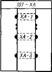
Fig. 17. Electropherogram showing how to cut off the edge of the sheet containing the separated fractions for micropreparative isolation.
The cut-off strip is stained; guided by it, the appropriate markings are made, and the fractions are eluted.
6. Qualitative Separation of amino acid mixtures. The separation and identification of all Amino Acids in complete acid hydrolysates of Proteins AND PEPTIDES cannot be achieved using a single paper electrophoresis method. For example, it is very difficult to identify both acidic (glutamic, aspartic, and cysteic) and basic (Histidine, Arginine, and Lysine) amino acids under the same conditions. At pH 5.0, good separation of glutamic, aspartic, and cysteic acids can be obtained, but under these conditions, basic amino acids fractionate poorly, and neutral Amino acids are obviously not separated at all, since at this pH their molecular charge is close to zero.
At the same time, basic amino acids, due to their different pK values, fractionate quite satisfactorily in a buffer solution at pH 6.5, since under these conditions histidine is only slightly protonated. Acidic amino acids, conversely, resolve poorly at pH 6.5. However, acidic and basic Amino acids can be separated simultaneously on a single electrophoregram using electrophoresis in a double-buffer system [3]. A sheet of filter paper is divided into two halves by a starting line. The cathodic half, which is the migration region for basic amino acids, is moistened with a pH 6.5 buffer solution, while the anodic half is impregnated with a pH 5.0 buffer solution (Fig. 18).

Fig. 18. One-dimensional paper electrophoresis.
1 — at pH 5.0; 2 — at pH 6.5; 3 — in a system with both buffer solutions.
Neutral amino acids do not separate in either of these buffer solutions, but their fractionation can be achieved by subsequent electrophoresis under different conditions, as well as by Chromatography. Transferring the neutral amino acid fraction to another system is very conveniently done by sewing (Fig. 19). For complete identification of amino acids, hydrolysates are applied to dry filter paper. The smaller the area occupied by the applied sample on the paper, the better the separation will be. Therefore, to minimize sample spreading, application is accompanied by drying. In the center of the paper sheet, a starting line is marked, and test samples 1, 2, and 3 are applied 3 cm apart (Fig. 19, A). On both sides of them, two control samples (K) are applied. In The First stage of the analysis, electrophoresis with two buffer solutions is performed. The cathodic part of the electrophoregram is moistened with a pH 6.5 buffer solution, and the anodic part with pH 5.0. Medium-voltage electrophoresis is run for about 1.5 hours, then the electrophoregram is removed from the apparatus, dried, and filter paper strips on which the control samples were fractionated are cut off from both sides. These strips 4 are stained with ninhydrin (Fig. 19, A). On the stained control electrophoregrams, the acidic and basic amino acids of the analyzed hydrolysates can be identified, and the localization of neutral amino acids can be established. From the original electrophoregram, a strip 5 containing only neutral amino acids is cut out along the dotted lines (Fig. 19, B). Both remaining parts of the sheet, designated by the number 6, are stained with ninhydrin, and the acidic and basic amino acids contained in them are identified by comparison with the control electrophoregrams. The paper strip containing the neutral amino acids is sewn to a new sheet of filter paper using a zigzag stitch on a sewing machine.

Fig. 19. Three-stage separation of amino acids.
A and B. Electrophoresis in a system with two buffer solutions. C. Electrophoresis at pH 1.9. D. Chromatography.
The next stage of electrophoresis is carried out in a buffer solution with a pH of 1.9 or 2.0. Therefore, the paper strip with neutral amino acids must be sewn onto the anodic edge of the sheet so that it serves as the starting zone. At pH 1.9–2.0, the dissociation of all carboxyl groups of amino acid molecules is suppressed, and consequently, all neutral Components of the mixture behave as cations. After sewing the paper strip, the sheet is turned over, and the area located beneath the sewn strip is carefully cut out with a razor blade. This must be done cautiously to avoid damaging the stitches and the strip itself. The filter paper is then moistened with a buffer solution having a pH of 1.9 or 2.0, as described above. As a marker, ε-DNP-lysine is applied next to the sewn strip. Electrophoresis is carried out until this marker has migrated 8–10 cm toward the cathode.
Upon completion of electrophoresis, the paper is dried again, one of the control strips 7 (Fig. 19, C) is cut out and stained. It helps determine how far the neutral Amino acids have migrated. Once this is established, the electrophoregram is cut (along the dotted lines) into strips containing the neutral amino acids from samples 1–3, as well as from the control sample K. Each of these strips is, in turn, sewn to a new sheet of filter paper and subjected to chromatography in a butanol–acetic acid–water system (Fig. 19, D) in a direction perpendicular to the direction of electrophoresis. Such a three-stage analysis system allows for the reliable identification of each amino acid contained in the analyzed hydrolysate.
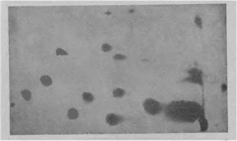
Fig. 20. Peptide map of the tryptic hydrolysate of D-glyceraldehyde-3-phosphate dehydrogenase.
Hydrolysates of short peptides containing only one or Two Types of amino acids can be analyzed using an abbreviated protocol, performing a two-stage instead of a three-stage separation.
7. Analysis of enzymatic protein hydrolysates. In 1957, Ingram [6] first demonstrated that complex peptide mixtures, enzymatic hydrolysates, etc., can be analyzed by a two-stage separation using paper chromatography and electrophoresis. Such an analysis reveals the most minor differences that may be encountered, for example, in species Specificity studies or other comparative research. The 'fingerprinting' method can be called one of the most important Methods in modern biochemistry, molecular biology, and immunochemistry, as it allows for the ISOLATION OF A specific peptide in pure form or the micropreparative separation of a complex mixture.
As a classic example of the 'fingerprinting' method, Fig. 20 shows the pattern obtained from the fractionation of a tryptic hydrolysate of D-glyceraldehyde-3-phosphate dehydrogenase.
In the first stage, horizontal electrophoresis is carried out at pH 4.3–5.0. 3–4 mg of the test material is applied to the paper in a 1 cm wide zone perpendicular to the direction of migration. Electrophoresis continues for 2–3 hours, after which the paper is dried and, turned by 90°, subjected to ascending chromatography. A mixture of isoamyl alcohol–pyridine–water (35 : 35 : 30) is used as the solvent. Chromatography is stopped when the solvent front reaches the upper edge of the paper. The chromatogram is dried and stained in the same manner as the electrophoregram in horizontal electrophoresis.

Fig. 21. Three-stage analysis of a protein hydrolysate.
A. Electrophoresis at pH 6.5. B. Electrophoresis at pH 1.9. C. Chromatography. See text.
Recently, the 'fingerprinting' method has been increasingly carried out in three stages, combining two electrophoretic separations with chromatography. The reason is that the two-stage 'fingerprinting' method does not always yield complete separation, especially when analyzing enzymatic hydrolysates of high-molecular-weight proteins; therefore, additional electrophoretic fractionation becomes necessary (Fig. 21).
In the first stage, it is best to perform electrophoresis in a buffer solution of pH 6.5 or 5.0, or in a double-buffer system. It is quite difficult to give specific recommendations regarding the pH of the buffer solution, as variations in electrophoretic mobility are much more significant for proteins and peptides than for amino acids. Therefore, in the first separation stage, the pH of the buffer solution should be selected empirically. It is also necessary to determine experimentally the most suitable Location for the starting zone and the optimal duration of electrophoresis, since the components of the mixture to be separated can have highly variable electrophoretic mobilities. Analysis of an unknown peptide mixture should begin with a preliminary electrophoretic run to establish these parameters.
After the first-stage electrophoretic separation, control strip 1 (Fig. 21, A) is stained, and using it as a reference, a strip 2 approximately 1 cm wide is cut from the unstained part of the paper sheet (Fig. 21, A). This strip is sewn onto a new sheet of paper for the second electrophoretic separation (Fig. 21, B), which is typically carried out at pH 2.1 or 1.9. At pH 1.9, complete separation of enzymatic protein hydrolysates cannot be expected due to the complex composition of zone 3 of neutral amino acids. Therefore, having established the location of this zone on the first electrophoregram, it is cut out and sewn next to strip 2 (Fig. 21, B). Electrophoresis continues until the ε-DNP-lysine applied as a marker has migrated approximately 10–13 cm toward the cathode. After drying, the paper is stained almost completely, except for strip 4 (Fig. 21, B), which contains the neutral components migrating from segment 3. Strip 4 is sewn to a new sheet and subjected to chromatography in an appropriate system (Fig. 21, C). Three-stage analysis allows for the most reliable Determination of the number of peptide components resulting from the Enzymatic Hydrolysis of high-molecular-weight proteins. Such data are extremely important, for example, in amino acid sequencing.
NOTES
1. The less buffer solution contained in the filter paper, the higher the resolution of horizontal electrophoresis. Therefore, before applying the sample, excess buffer solution should be removed particularly thoroughly by blotting.
2. If irregularly shaped zones with edge distortion are formed, or if the migration front is not parallel to the electrodes, the correct fit of the lid must be checked. Under an insufficiently pressed or warped lid, the paper becomes unevenly wetted because it dries out in some areas, which can cause such anomalies. If the lid is well secured but these phenomena persist, the correct placement of the cellophane strips should be checked. If the cellophane spacers are too short, the filter paper on one or both sides may come into direct contact with the wick. This will cause an increased flow of buffer solution at the point of contact and, consequently, distortion of the zone shapes. In high-voltage horizontal electrophoresis, The Effect of electroendosmosis is less pronounced, so cellophane spacers can be omitted.
3. In fingerprinting analysis, it is recommended to perform chromatography, as well as electrophoresis, at a low temperature, if possible. Our experience indicates that the highest resolution is observed at + 4°C.
Last update: 06/08/2026
Editorial and Educational Adaptation: This material has been compiled based on the primary/original source text. The project team performed an editorial review, corrected technical inaccuracies, structured sections, and adapted the content for an educational format.
What was processed:
- elimination of formatting defects (OCR errors, structural breaks, corrupted characters);
- editorial organization of content;
- standardization of terminology in accordance with academic sources;
- verification of factual statements against the original source text.
All mentions of the author, publication year, and origin of the primary text have been preserved in accordance with the source.