BIOTECHNOLOGY - V. H. Gerasymenko - 2006
Part I. General Biotechnology
CHAPTER 4. CELL ENGINEERING
4.5. BIOTECHNOLOGY OF GENE TRANSFER INTO EUKARYOTIC CELLS VIA DNA (DNA TECHNOLOGY)
For Gene transfer into somatic Cells, both total DNA obtained by purifying DNA preparations from salmon sperm or cells lines (most commonly of mouse, Chinese hamster, and human origin) and cloned genes are used. When transforming somatic cells growing in culture and mutant lines derived from Cell cultures using total DNA preparations and cloned genes, TK, HPRT, APRT, and DHFR are most frequently used as donor marker genes; they encode the Enzymes TK (thymidine kinase), HPRT (hypoxanthine-guanine phosphoribosyltransferase), APRT (adenine phosphoribosyltransferase), and DHFR (Dihydrofolate Reductase), respectively. Thus, the transformation of mammalian cells in culture requires a source of donor DNA, a suitably buffered medium, marked recipient cells, and/or a suitable selective medium for culturing transformants.
When a cell culture is treated with exogenous DNA preparations, approximately one in a million or one in 100 million (10-6-10-8) recipient cells becomes transformed. At the same time, It is worth noting that the transformation frequency of somatic recipient cells using metaphase Chromosomes as vectors was generally one to two orders of magnitude higher. Achieving a high frequency of somatic cell transformation with exogenous DNA relies crucially on the source of high-molecular-weight carrier DNA, a properly buffered medium, and the physiological state of freshly seeded recipient cells.
In transformation experiments, somatic cell-derived DNA with a size exceeding 100 kb is typically used as the donor, with an optimal concentration considered to be 500 mcg/ml.
To enhance transformation efficiency, Serov O. L. (1985) suggested treating purified viral DNA preparations with DEAE-dextran, which increases the resistance of the DNA to thermal Denaturation and the hydrolytic action of endogenous DNases present in the recipient cell. Ultimately, this leads to increased transformation efficiency. In recent years, a hundredfold increase in transformation efficiency has been achieved by treating cultured somatic recipient cells with a DNA-calcium phosphate complex (precipitate). Transformation success depends on the reaction medium during the precipitation of exogenous DNA with calcium phosphate, the concentration of DNA used to form the complex, the Treatment of recipient cells with dimethyl sulfoxide, and the duration of incubation of cultured somatic recipient cells with the DNA-calcium phosphate precipitate. Some of these parameters do not have a universal transforming effect, but rather increase the number of transformed recipient cells only with respect to specific cell lines.
Selection schemes for recessive and dominant genes differ. The DNA-calcium phosphate precipitate enters the recipient cell through adsorption of the complex onto The Cell membrane surface and active phagocytosis, which occurs During the first hours of incubation of the DNA-calcium phosphate precipitate with the recipient cells. Furthermore, it has been established that adsorption and phagocytosis are energy-dependent processes and are suppressed when cellular Respiration is inhibited. Within the first hour of incubation, the DNA-calcium phosphate precipitate is found on the cytoplasmic membrane, then in cytoplasmic vacuoles, and 8 hours after THE START OF incubation, a small portion of the exogenous donor DNA reaches the Nucleus of the cultured recipient cells. At all stages of cell entry, the exogenous DNA remains bound to the calcium phosphate precipitate, and the Size and Structure of the donor DNA within the recipient cell are preserved for 24 hours from the start of incubation.
Gene transfer into recipient somatic cells as part of total DNA is possible using carriers such as Liposomes (phospholipid vesicles) or erythrocyte ghosts, into which the isolated nucleic acid preparation has been pre-encapsulated by one method or another. This method yields a higher number of transformed somatic cells than DNA transfer via other Methods. In addition, transforming recipient cells with total DNA preparations pre-encapsulated in liposomes is relatively straightforward.
Liposomes possess low toxicity and are capable of protecting encapsulated Nucleic Acids and other macromolecules from degradation. There are no significant limitations on producing liposomes of sizes that allow the encapsulation of Biopolymers spanning a wide molecular weight range (from purified genes to chromosomes). To ensure selective affinity for specific recipient cells and increase binding capacity to the membranes of particular cell populations, the liposome surface can be modified using Glycolipids, Lectins, or covalently linked Antibodies. It is suggested that using liposomes for DNA delivery enhances somatic cell transformation efficiency and enables transformation studies in cells for which other DNA transfer methods are unsuitable.
Among the known methods of liposome preparation, the reverse-phase evaporation method is the most suitable for transferring total DNA into recipient somatic cells, allowing DNA with a mass of 108 daltons to be incorporated into liposomes. To increase the efficiency of liposome-mediated DNA transfer into somatic cells, dimethyl sulfoxide, Ethylene glycol, glycerol, and polyethylene glycol are used. The last of these substances increases the transforming capacity of liposome-encapsulated DNA by 10-fold, although moderate concentrations of glycerol, in which cells are maintained after contact with liposomes, are even more effective. Evidence indicates that exogenous SV40 viral DNA placed in negatively charged liposomes appears up to three orders of magnitude more infectious than when encapsulated in neutral liposomes. Regarding process dynamics, the most efficient transformation of somatic cells with liposome-encapsulated DNA occurs during the first 30 minutes of incubation. There are also Examples demonstrating high transformation efficiency in plant protoplasts using purified Ti-plasmid DNA encapsulated in liposomes.
In recent years, research has successfully focused on the competence for transformation of cultured recipient somatic cells treated with exogenous DNA preparations. Compelling evidence points, on the one hand, to the genetic nature of high cellular competence for transformation, and, on the other hand, demonstrates that the efficiency of exogenous DNA transfer depends on the physiological state of the recipient cells, their tissue origin, and their differentiation.
Transfer of cloned and purified genes into somatic cells. When studying the transformation of somatic and Germ Cells, O. L. Serov most frequently used the cloned Herpes simplex virus (Herpes simplex) TK gene, treating viral genomic DNA with the Restriction Endonucleases HpaI and BamHI. The resulting DNA fragments obtained through Enzymatic Hydrolysis, containing 3.3 and 3.4 kilobase pairs respectively, harbor the TK gene; the mRNA transcribed from the intact herpes virus TK gene is a sequence consisting of nearly 1.3 thousand nucleotide residues. Furthermore, it was established that during thymidine kinase Biosynthesis, a 107-nucleotide region starting from the 5' end of the mRNA is not translated. Fragments of viral genomic DNA containing the TK gene are cloned using Plasmids and phages as vectors.
Transformation of somatic and germ cells with the TK gene is carried out using recombinant DNA constructed by inserting herpes virus DNA fragments containing the thymidine kinase-encoding gene. To increase transformation efficiency, somatic or germ cells are treated with purified or cloned genes via calcium phosphate-mediated donor DNA precipitation. This approach successfully introduces a purified or cloned gene into one out of every 105-107 treated recipient cells.
A more efficient gene transfer method compared to those described above is the microinjection of DNA in solution directly into the nuclei of recipient cells. In this case, 50-100% of the treated cells are transformed. A similar transformation frequency is achieved even when The Nucleus is merely punctured with a microneedle. The DNA representing the purified or cloned gene is not directly injected into the nucleus in this instance, but is instead used to treat cultured mammalian cells. Other methods for transferring cloned or purified genes into mammalian somatic recipient cells include treating recipient cells with liposome-encapsulated DNA preparations or high concentrations of polyethylene glycol; the latter achieves a transformation frequency comparable to that attained using the nuclear microinjection method for DNA solutions.
Attention should also be drawn to cotransformation, a method gaining widespread use for introducing foreign unselected genes or DNA fragments into recipient cells. A method of transforming recipient cells using electrical discharges in a high-voltage field is also under development.
When a purified or cloned gene encoding thymidine kinase is used as the donor for transformation, cultured mutant cells lacking the TK gene in their genome serve as recipients, and HAT selective medium is used to separate transformed from untransformed cells (Fig. 4.3). Recipient cells transformed with the TK gene are capable of growing on HAT medium, whereas untransformed cells perish. The presence or absence of carrier DNA has a significant impact on the transfer frequency of the cloned herpes virus TK gene into eukaryotic recipient cells. Transformation of Chinese hamster recipient cells with the cloned herpes virus TK gene in the absence of carrier DNA was more than 200 times lower compared to experiments in which the cloned herpes virus TK gene was transferred into recipient cells with the assistance of carrier DNA. Researchers believe The Role of carrier DNA is to protect the transferred cloned or purified gene from the degrading action of hydrolytic enzymes within the recipient cells. The success of introducing purified or cloned genes into somatic cells via the calcium phosphate method depends largely on the concentration of donor DNA. Although many mechanisms by which carrier DNA contributes to the successful transformation of recipient cells with purified or cloned genetic material—as well as the role of carrier DNA in transgenome formation—remain unclear, the structural alteration of purified or cloned genes introduced into recipient cells in the presence of carrier DNA is considered a firmly established fact. It is also likely established that structural changes in exogenous cloned genetic material during transgenome formation are observed only when carrier DNA enters the recipient cell simultaneously. The role of the carrier DNA is to protect the selected genes from subsequent degradation by incorporating the selected genetic material into the carrier Introduction/20.html">DNA Structure, thereby creating a transgenome with novel properties. In the absence of carrier DNA, selected genes that have undergone structural changes, predominantly driven by the enzymatic activity of the recipient cells, are eliminated, while intact introduced purified or cloned genes remain within the recipient cell.
Class="center">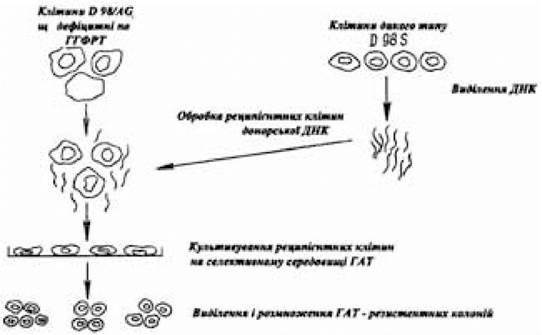
Fig. 4.3. Schematic diagram of somatic cell transformation experiments using DNA
(after Shybalska Ye. and Shybalskyi V., 1982)
Consequently, transformed cells may simultaneously harbor intact exogenous DNA molecules and DNA molecules that have undergone structural changes. Recipient cells transformed using total DNA exhibit a phenotype that may display stable or unstable expression. Evidence indicates that stabilization of the transformed phenotype results from donor gene Amplification or the integration of this gene into the recipient cell genome, although mechanisms opposing the integration of exogenous DNA into the recipient cell genome may also operate.
The appearance of stable transformants has been observed upon the integration of donor genetic material, including cloned gene DNA, into the chromosomes of recipient cells, which leads to the destabilization of the latter and the subsequent emergence of numerous Chromosomal aberrations. In stable transformants formed As a result of the integration of donor genes introduced into the recipient genomic material via total DNA as well as purified or cloned genes, a process of destabilization occurs due to their inherent instability. This process is not always the result of losing the integrated donor gene; sometimes the expression of the donor gene is altered, preventing the realization of the transformed phenotype.
A clear correlation is observed between the state of the transgenome (stable vs. unstable), the frequency of donor genetic material integration into the recipient cell genome, and the frequency of transgenome stability disruption in stable transformants, on the one hand, and the degree of differentiation and karyotype status of the recipient cells, on the other. Thus, recipient cell lines with altered heteroploid karyotypes (fibroblast-like cells) are capable of retaining autonomously replicating donor genetic material in their genomes with an unstable transformed phenotype, and destabilizing the transformed phenotype in stable clones. Conversely, recipient cells with a near-normal or normal karyotype (germ cells) lack the conditions that favor the transgenome remaining in an unstable autonomous state. Therefore, a high proportion of stable transformants is observed during the transformation of recipient germ cells with exogenous DNA.
Last update: 11/08/2026
Editorial and Educational Adaptation: This material has been compiled based on the primary/original source text. The project team performed an editorial review, corrected technical inaccuracies, structured sections, and adapted the content for an educational format.
What was processed:
- elimination of formatting defects (OCR errors, structural breaks, corrupted characters);
- editorial organization of content;
- standardization of terminology in accordance with academic sources;
- verification of factual statements against the original source text.
All mentions of the author, publication year, and origin of the primary text have been preserved in accordance with the source.