Diagnosis and treatment of patients with recurrent gastroduodenal bleeding - Shaprynskyi V.O. 2009
Surgical treatment of recurrent ulcerative gastroduodenal hemorrhage
Vagotomy options for recurrent bleeding duodenal ulcers
TRUNCAL VAGOTOMY.
Transection of the anterior trunk of the n.vagus denervates the anterior PARTS OF THE Stomach and simultaneously leads to denervation of the Liver and biliary tract. Transection of the posterior trunk completes the denervation of The Stomach and also denervates the Pancreas and intestines. Therefore, it is not surprising that total subdiaphragmatic (truncal) vagotomy leads to such undesirable consequences as impaired MOTOR FUNCTION OF the Gallbladder and Bile ducts, diarrhea, and pancreatic dysfunction. At the same time, complete denervation of the stomach, along with a reduction in its acid-producing function, leads to significant gastric motor and emptying disorders (Fig. 10.3.1).
In this operation, after performing an upper midline laparotomy, traction is applied to the stomach downward and to the left, and its fundus is retracted laterally. After these steps, visual access to the abdominal Esophagus is obtained. Beneath the Peritoneum along the anterior surface of the esophagus, the anterior trunk of the n.vagus is put on tension, transected, and resected over a length of 1-2 cm. The posterior trunk of the n.vagus is located by passing a finger around the esophagus, and is also transected and resected over a length of 1-2 cm (Figs. 10.3.2-6). Afterwards, the integrity of the peritoneum in the region of the abdominal esophagus is restored.
In some cases, it is difficult to locate the Vagus nerve trunks using these maneuvers, so additional mobilization of the left lobe of the liver is required. To do this, the left triangular ligament of the liver must be transected along its avascular zone. The incision is made 5-6 cm long, closer to the liver to avoid damaging the phrenic vein. To facilitate gastric traction in such cases, a sling is passed around the esophagus. To provide a reliable landmark in the esophageal region, the insertion of a thick nasogastric tube is advisable. Slightly above the esophageal hiatus of the Diaphragm, a 4 cm incision is made in the peritoneal sheet (transecting the phrenicoesophageal ligament). After digital exploration of the esophagus, the anterior vagal trunk and its accessory branches are easily found. By transecting the gastrocolic ligament and applying downward traction on the posterior wall of the stomach, the posterior trunk of the n.vagus can be located more quickly; it is palpated as a cord between the esophagus, the right crus of the diaphragm, and the aorta. Sometimes it is easier to detect by passing a finger around the esophagus from left to right. In some cases, when the celiac branch is clearly identified, the main posterior (right) vagal trunk can be found by advancing a finger deep along the course of this branch and applying slight tension to it.
SELECTIVE VAGOTOMY.
This type of surgical intervention consists of transecting all gastric fibers of the anterior and posterior trunks of the n.vagus. At the same time, functionally important branches going to The Liver and celiac plexus are preserved. Selective vagotomy is recommended as a means of reducing the incidence of dumping syndrome by preserving the vagal Innervation of the liver and Small Intestine. There are two variants of selective vagotomy: 1) transection of the gastric Branches of the n.vagus without isolating the main trunks and with ligation of the a.gastrica sinistra (Griffits, 1966; Pantsyrev, Grinberg, 1979) and 2) with Isolation of the main trunks of the vagus nerve and transection of the gastric branches while preserving the a.gastrica sinistra (Burge, 1964; Tanner, 1965; Shalimov, 1975) (Fig. 10.3.7).
Class="center">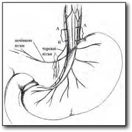
Fig. 10.3.1. Schematic of truncal vagotomy.
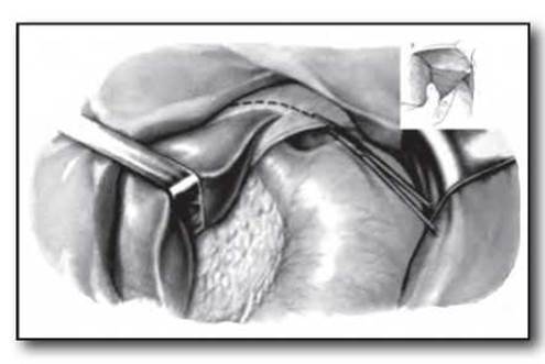
Fig. 10.3.2. Stages of truncal vagotomy. Transection of the left triangular ligament of the liver.
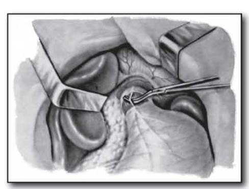
Fig. 10.3.3. Stages of truncal vagotomy. Isolation of the anterior trunk of the vagus nerve.
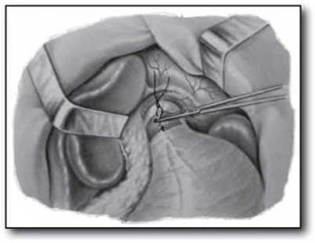
Fig. 10.3.4. Stages of truncal vagotomy. Transection and ligation of the anterior trunk of the vagus nerve.
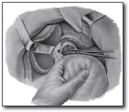
Fig. 10.3.5. Stages of truncal vagotomy. Isolation of the posterior trunk of the vagus nerve.
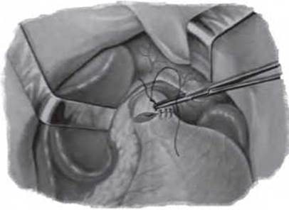
Fig. 10.3.6. Stages of truncal vagotomy. Restoration of peritoneal integrity in the region of the abdominal esophagus.
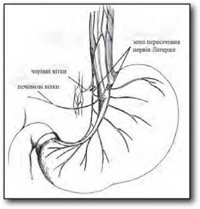
Fig. 10.3.7. Schematic of selective vagotomy.
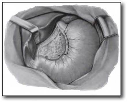
Fig. 10.3.8. Stages of selective vagotomy.
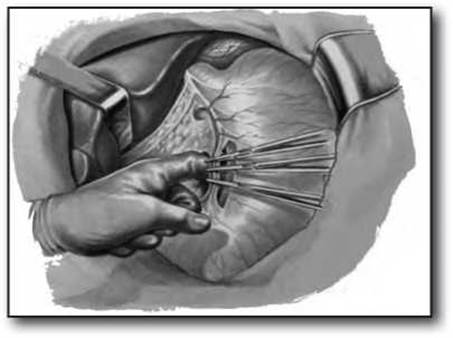
Fig. 10.3.9. Stages of selective vagotomy.
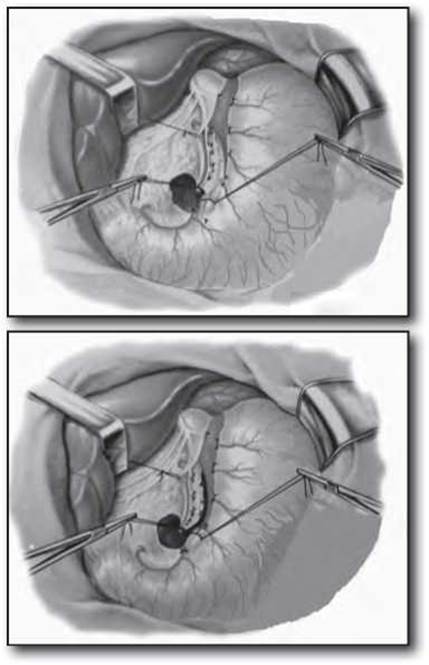
Figs. 10.3.10 and 10.3.11.
Stages of selective vagotomy. Transection and ligation of the anterior and posterior leaflets of the gastric serosa in the region of the lesser omentum.
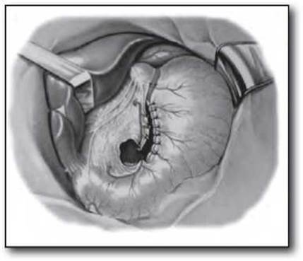
Fig. 10.3.12. Stages of selective vagotomy. Restoration of the integrity of the gastric serosa.
Transection of the gastric branches of the vagus nerve without isolating the main trunks and with ligation of the a. gastrica sinistra. After verifying the anterior trunk of the n. vagus, we identify its main gastric branch, the so-called nerve of Latarjet, which is palpated along the lesser curvature of the gastric body and is covered by the peritoneum of the lesser omentum. In the uppermost part of the n. vagus, the hepatic branch of the trunk is clearly identified, which branches off to the right and runs toward the porta hepatis. Often, there may be two or three such branches. An accessory hepatic artery, arising directly from the a. gastrica sinistra, may run parallel to the hepatic branch of the n. vagus. The avascular portion of the lesser omentum is divided with scissors, and the a. gastrica sinistra is verified, clamped with three hemostatic forceps, divided, and ligated. Along the planned line from the lesser curvature to the left margin of the cardia (angle of His), the gastric serosa is incised and ligated in separate bundles. By transecting all gastric branches of the anterior trunk of the n. vagus and their accompanying vessels, the posterior leaflet of the lesser omentum, along with the vessels and branches of the posterior vagal trunk, is exposed. The stump of the a. gastrica sinistra is retracted to the right, while the pars nuda of the lesser curvature of the stomach, previously secured with a traction suture, is retracted to the left. All gastric branches of the posterior vagal trunk and their accompanying vessels are transected and ligated. Additionally, the branches of the n. vagus arising below the esophageal hiatus of the diaphragm are transected and ligated. The lesser curvature of the stomach is peritonized with interrupted sero-serosal sutures (Figs. 10.3.8-12).
Transection of the gastric branches of the vagus nerve with isolation of the main vagal trunks and division of the gastric branches while preserving the a. gastrica sinistra. After verifying the anterior (left) trunk of the vagus nerve, we place it on a traction suture. We apply traction to the stomach downward and to the left, and to the anterior trunk of the n. vagus via the traction suture to the right, after which the gastric and hepatic branches are clearly identified. Between the esophagus and the right crus of the diaphragm, we locate the posterior (right) vagus nerve, which is slightly elevated while simultaneously applying downward and leftward traction to the stomach. The surgeon elevates the left gastric artery on the index finger and identifies the triangle formed by the a. gastrica sinistra, the celiac branch, and the posterior trunk of the n. vagus. We transect the trunk of the vagus nerve below the Water/144.html">Origin of the celiac branch to the solar plexus, ensuring the transection of the branches running from the trunk to the gastric cardia and esophagus.
SELECTIVE PROXIMAL VAGOTOMY.
Selective vagotomy of the gastric parietal Cell mass (highly selective vagotomy) is a partial denervation of the stomach within its body and fundus (i.e., the regions where the mucosa contains the acid-producing zone). We isolate the trunks of the anterior and posterior n. vagus, placing them on separate traction sutures (Fig. 10.3.13).
Following the aforementioned maneuvers, we determine the boundary for the transection of the vagal nerve branches, which should coincide with the junction between the antrum and the body of the stomach. To identify this boundary, we apply downward traction to the stomach and locate the anterior nerve of Latarjet, which typically enters the gastric wall 6-7 cm from the pylorus in the form of a "crow's FOOT". The proximal branch of this "crow's foot" corresponds to the boundary between the antrum and the body of the stomach along the lesser curvature. Along the greater curvature, this boundary is usually at the same distance from the pylorus as along the lesser curvature and coincides with THE ORIGIN OF the first gastric branch of the a. gastroepiploica dextra. Additionally, intragastric pH-metry should be utilized. Through the mobilized area of the stomach along the greater curvature in the region of the gastrocolic ligament, we inspect the posterior surface of the lesser omentum. We locate the Termination of the posterior branch of the nerve of Latarjet and determine the starting point for the transection of the gastric branches of this nerve.
Having established the starting point of the vagotomy, we transect and ligate several neurovascular bundles along with portions of the anterior and posterior peritoneal leaflets of the lesser omentum right at the edge of the lesser curvature of the stomach. Two traction sutures are passed through the created opening. Gentle traction on the gastric sutures downward and to the left, and on the lesser omentum upward and to the right, facilitates the further dissection of the lesser omentum from the stomach.
First, the anterior leaflet of the peritoneum of the lesser omentum is dissected by transecting and ligating the neurovascular bundles in small portions. Upon reaching the cardia, the direction of transection is changed, dividing the peritoneum in the cardiac region along with the Vessels and nerves up to the angle of His. The esophagus is then mobilized for at least 5 - 6 cm above the cardia.
The esophagocardiac branches of the n. vagus, which arise from the anterior vagal trunk, are intimately adherent to the anterior wall of the esophagus. They must be carefully transected while holding
the main trunk of the n. vagus by the previously placed traction suture.
Through the gradual, step-by-step transection of the neurovascular bundles along with the posterior peritoneal leaflet up to the cardia, the lesser curvature of the stomach is completely freed from the lesser omentum.
To facilitate exposure and denervation of the posterior aspect of the cardia and esophagus, a traction tape is placed at this level. Applying traction to the tape placed on the posterior trunk of the n. vagus and on the esophagus creates favorable conditions for transecting the numerous nerve branches running to the cardia and esophagus, skeletonizing the latter for at least 5 - 6 cm above the cardia. To facilitate this step, two fingers of the left hand can be inserted as retractors between the lesser curvature on the left and the nerve of Latarjet on the right. The integrity of the nerve of Latarjet is verified by periodic traction on the main trunk via the traction suture. The phrenicogastric ligament is sequentially transected up to the first short gastric vessels, ligating the first of them. For more complete denervation, a circular myotomy of the longitudinal Muscle layer of the esophagus is performed 1.5 - 2 cm above the cardia.
An important point is that the terminal branches of the nerve of Latarjet run both to the antrum and as recurrent branches upward to the lesser curvature of the gastric body. Therefore, to achieve complete denervation of the gastric body while preserving the innervation of the antrum, O. O. Shalimov proposed performing a transverse incision of all gastric layers down to the mucosa above the "crow's foot," extending onto the anterior and posterior walls of the stomach by 1.5 - 2 cm, after which the edges of the incisions are sutured longitudinally.
In addition to this, along the greater curvature of the stomach at the boundary between the antrum and the body, O. O. Shalimov proposed transecting the a. gastroepiploica dextra along with its accompanying nerve trunks (Figs. 10.3.14-19).
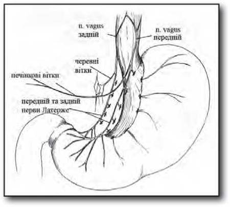
Fig. 10.3.13. Schematic of selective proximal vagotomy.
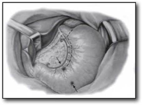
Fig. 10.3.14. Stages of selective proximal vagotomy. Determining the boundary for the transection of the n. vagus nerve branches.
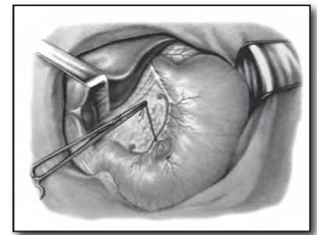
Fig. 10.3.15. Stages of selective proximal vagotomy. Transection and ligation of neurovascular bundles along with portions of the anterior and posterior peritoneal leaflets of the lesser omentum.
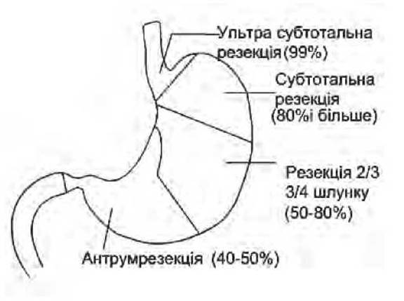
Fig. 10.4.1. Level and percentage of gastric resection in its various types.
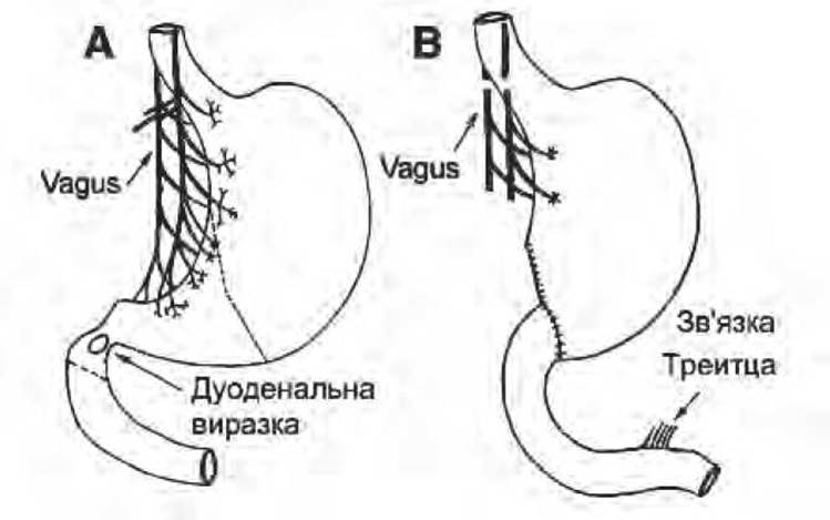
Fig. 10.4.2. Antrectomy with selective bilateral vagotomy and Billroth I restoration of gastrointestinal tract continuity.
Last update: 11/08/2026
Editorial and Educational Adaptation: This material has been compiled based on the primary/original source text. The project team performed an editorial review, corrected technical inaccuracies, structured sections, and adapted the content for an educational format.
What was processed:
- elimination of formatting defects (OCR errors, structural breaks, corrupted characters);
- editorial organization of content;
- standardization of terminology in accordance with academic sources;
- verification of factual statements against the original source text.
All mentions of the author, publication year, and origin of the primary text have been preserved in accordance with the source.