Diagnosis and treatment of patients with recurrent gastroduodenal hemorrhage - Shaprynskyi V.O. 2009
Surgical treatment of recurrent bleeding gastroduodenal ulcers
Types of pyloroduodenoplasty for acutely bleeding duodenal ulcers
In the surgery of acutely bleeding ulcers, a pathogenetically substantiated surgical intervention is ulcer excision followed by pyloroduodenal reconstruction and one of the types of vagotomy. Vagotomy supplemented by pyloroplasty as a standalone Procedure is not justified for this pathology. Therefore, in this section of the book, we will discuss the possible types of pyloroduodenal reconstruction after ulcer excision.
All modern modifications of pyloroduodenal reconstruction are based on three classic Methods developed in the late 19th and early 20th centuries.
In 1886, Heineke, and in 1888, Mikulicz proposed a pyloroplasty method to manage pyloroduodenal stenosis, which involved a 6-8 cm longitudinal incision through the pylorus and the duodenal bulb along the axis of the pyloric region. The longer part, namely 2/3 of the incision, was made on the Cytology/practical/109.html">Pyloric part of The Stomach, while the shorter part, about 1/3 of the incision, extended toward the duodenum. The landmark for identifying the pylorus is the so-called pyloric vein of Mayo. To facilitate the surgeon's maneuvers in this area, it is advisable to perform a preliminary Kocher mobilization of the duodenum. After incising the pyloric and duodenal lumen, their walls were tractioned transversely relative to the axis of the pyloric canal using stay sutures and closed transversely with a double-row suture (Fig. 10.2.1).
The most commonly used method is the Judd (1915)-Horsley (1919) pyloroplasty, which consists of a diamond-shaped excision of the ulcer tissue on the anterior duodenal wall. Subsequently, this technique was modified by using an oval (Judd-Horsley) or rectangular (Judd-Tanaka) excision of the ulcer tissue. However, regardless of the excision variant used for the anterior wall ulcer, the procedure was completed with a Heineke-Mikulicz pyloroplasty (Figs. 10.2.2; 10.2.3).
Class="center">
Fig. 10.2.1. Stages of Heineke-Mikulicz pyloroplasty (a, b, c)
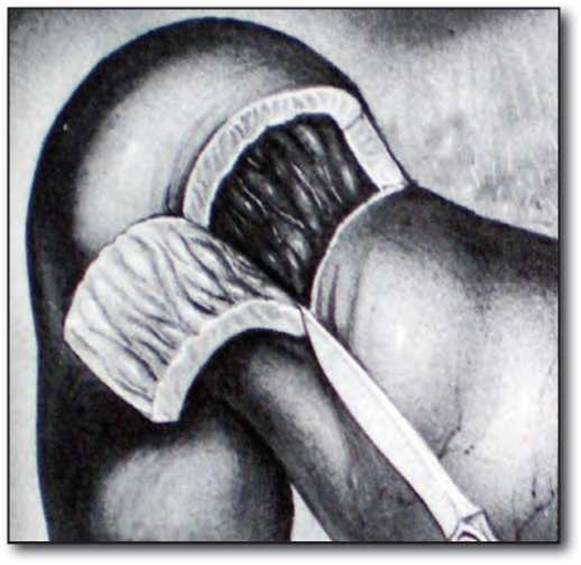
Fig. 10.2.2. Judd-Tanaka pyloroplasty.

Fig. 10.2.3. Judd-Horsley pyloroplasty.

Fig. 10.2.4. Stages of Finney pyloroplasty (a-e)

Fig. 10.2.5. Stages of Mayo pyloroplasty (a-b)
In 1902, Finney proposed a horseshoe-shaped (U-shaped) incision of the pyloroduodenal zone, followed by suturing the inner and outer edges of the incision with a double-row suture. Before performing the pyloroplasty, it is crucial to divide all adhesions and perform an extended Kocher mobilization of the duodenum along with mobilization of the pyloric region of the stomach and the pylorus. The first stay suture is placed at the upper edge of the mid-pylorus, and the second is placed 4-5 cm distally on the duodenal wall and proximally on the wall of the pyloric region of the stomach, bringing them together and forming the posterior wall of the pyloroduodenal anastomosis using seroserous sutures. A U-shaped incision of the pylorus and duodenum is performed, followed by closure with a continuous suture and the placement of a second row of seroserous sutures on the anterior wall of the anastomosis (Fig. 10.2.4).
In 1954, Mayo proposed the excision of a stenosing pylorobulbar ulcer followed by a Finney-type pyloroplasty (Fig. 10.2.5).
After vagotomy, one type of gastric drainage procedure is the Deaver-Burden (1929)-Shalimov (1965) pyloroplasty. The pylorus is delivered into the wound, and two stay sutures are placed on its anterior surface 3 cm apart, after which an incision is made down to the muscular layer of the pylorus. Using partly sharp and partly blunt dissection, the muscular sphincter is isolated and excised over a length of about 2 cm. The mucosa, which we attempt to keep intact, bulges prominently at the site of the muscular sphincter defect. The incised seromuscular wall is then sutured in a transverse direction. The advantage of this technique is that it does not shorten the stomach along its axis, nor does it open the lumen of the gastrointestinal tract (Fig. 10.2.6).
In 1892, Jabouley proposed managing pyloroduodenal stenosis by creating an anastomosis between the pyloric region of the stomach and the anteromedial wall of the duodenum below the site of the stenosis. However, this method of gastric drainage has currently been abandoned.
These classic pyloroplasty techniques laid the foundation for more modern duodenoplasty methods, i.e., reconstruction of the duodenum without dividing the pyloric sphincter. Thus, in 1971, Tanner proposed that in cases of stenotic deformity of the duodenal bulb with uniform prestenotic dilation, a longitudinal incision should be made along the axis of the duodenum, followed by suturing the walls in the fashion of a Heineke-Mikulicz pyloroplasty.
In 1972, Kennedy proposed a horseshoe-shaped widening of the stenosis followed by a duodenoplasty in the fashion of a Finney pyloroplasty in cases with large prestenotic dilation of the duodenal bulb. However, despite the organ-preserving nature of these Procedures—namely, the maximum preservation of the pyloric sphincter—the Tanner-Kennedy operations remained palliative, as they did not involve excision of the ulcer tissue (Fig. 10.2.7).
In 1981, Barroso developed his own duodenoplasty technique, which consisted of a transverse excision of the stenosing duodenal ring en bloc with the ulcer tissue, followed by a widening closure of the walls in a transverse direction (Fig. 10.2.8).

Fig. 10.2.6. Stages of Deaver-Burden-Shalimov pyloroplasty (a-c).

Fig. 10.2.7. Stages of Tanner-Kennedy pyloroplasty (a-c).

Fig. 10.2.8. Stages of Barosso pyloroplasty (a-c).

Fig. 10.2.9. Stages of Helwing and Heyman pyloroplasty (a-e).
To address The Challenge of removing callous, deeply penetrating, and concurrently stenosing ulcers, Helwing and Heyman in 1978 proposed prepyloric segmental resection of the duodenum with exteriotorization of the ulcer crater beyond the gastrointestinal lumen, which was an undeniable advantage of this procedure at the time of its development. The ulcer crater is left on the HEAD of the Pancreas or the hepatoduodenal ligament, and the vessel at the Base of the ulcer is transfixed with interrupted sutures. A duodenal segment is resected using two parallel incisions corresponding to the width of the penetrating ulcer crater. The distal end of the posterior duodenal wall is not separated from the crater, while the proximal end of the wall is sutured to the distal one. Consequently, the ulcer crater is tamponaded by the proximal portion of the posterior duodenal wall. This represents a major drawback of the procedure, as suturing the posterior wall is performed virtually on the callous margin of the ulcer, posing a high risk of anastomotic leakage. The Helwing–Heyman procedure can hardly be classified as duodenoplasty; rather, it is a radical circular duodenal resection with exclusion of the ulcer from the gastrointestinal lumen (Fig. 10.2.9).
In 1982, V.T. Zaitsev and M.M. Velihotskyi developed new Methods for the radical excision of duodenal and pyloric ulcers complicated by bleeding. The authors described three methods of duodenoplasty: segmental, subcircular, and circular.
When the ulcer is localized on the superior, posterosuperior, inferior, or posteroinferior wall of the duodenum, penetrating into the head of the pancreas and the hepatoduodenal ligament, mobilization of the pylorobulbar zone is performed. If the ulcer is located on the posterosuperior wall, the right gastric artery (a. gastrica dextra) is often ligated. To facilitate the excision or exteriotorization of the ulcer, a stay suture can be placed on the posterior duodenal wall, and traction can be applied to pull it forward. After excision of the ulcer along with the surrounding scar tissue and areas of penetration (which are excluded from the gastrointestinal lumen), reconstruction of the duodenal lumen begins from the posterior wall in a transverse direction relative to the longitudinal axis of the duodenum. The authors termed this method of restoring duodenal integrity as arcuate segmental duodenoplasty (Fig. 10.2.10).
In cases of giant ulcers or double duodenal ulcers, especially kissing (mirror) ulcers penetrating the posterosuperior and posteroinferior walls, a small bridge of the intestinal wall remains after excision and exteriotorization of these ulcers, which comprise 3/4 of the duodenal circumference. Reconstruction of the intestinal lumen begins at the posterior wall, extending through the inferior and superior walls to the anterior wall. The authors termed this type of duodenoplasty subcircular (Fig. 10.2.11).
In 1980–1984, V.I. Onopriev referred to segmental and subcircular duodenoplasty by the single term 'bridge duodenoplasty'.
In the case of circular ulceration of the duodenal wall, the authors propose complete transection of the duodenal lumen with excision or exteriotorization of the ulcer, followed by end-to-end duodenoduodenostomy (circular duodenoplasty) (Fig. 10.2.12).
Ulcers located in the postbulbar region present a particular challenge. If the ulcer is parapapillary, reconstruction of the defect can be performed using the lateral and anterior walls of the duodenum. In cases where the ulcer process stenoses the major duodenal papilla, the ulcer is excised along with the stenotic part of the papilla, combined with concurrent papillosphincteroplasty. In the presence of a Wirsung's duct stricture, papillosphincteroplasty should be supplemented with wirsungoplasty (Fig. 10.2.13).


Fig. 10.2.10. Stages of segmental duodenoplasty (a-c).

Fig. 10.2.11. Subcircular duodenoplasty.

Fig. 10.2.12. Stages of circular duodenoplasty (a-b)

Fig. 10.2.13. Papillosphincteroplasty, wirsungoplasty.
When a choledochoduodenal fistula is diagnosed, duodenoplasty should be supplemented with choledochoduodenostomy.
Last update: 11/08/2026
Editorial and Educational Adaptation: This material has been compiled based on the primary/original source text. The project team performed an editorial review, corrected technical inaccuracies, structured sections, and adapted the content for an educational format.
What was processed:
- elimination of formatting defects (OCR errors, structural breaks, corrupted characters);
- editorial organization of content;
- standardization of terminology in accordance with academic sources;
- verification of factual statements against the original source text.
All mentions of the author, publication year, and origin of the primary text have been preserved in accordance with the source.