Review of Medical Physiology - William F. Ganong 2002
Endocrine System, Metabolism, and Reproduction
Pituitary Gland
Physiology of Growth
Growth is a complex phenomenon influenced not only by Growth Hormone and somatomedins, but also by THYROID Hormones, androgens, estrogens, glucocorticoids, and Insulin. In addition, normal growth obviously depends on genetic factors and adequate Nutrition. Normally, it involves a sequential series of changes during maturation, resulting in an increase in protein mass and overall linear dimensions, as well as weight gain that may stem from the accumulation of adipose tissue or salt and Water retention.
The Role of Nutrition
Food is the primary external factor affecting growth. Nutrition must be well-balanced—not only in protein content, but also in essential Vitamins, minerals (see Chapter 17), and caloric intake, ensuring that ingested protein is not simply burned for energy. The age at which nutritional deficiency occurs is also critical. For instance, rapid linear growth continues during Puberty even when caloric intake is restricted. Trauma and illness stunt growth by accelerating Protein Catabolism.
Periods of Growth
The growth process varies across species. Rats grow throughout their entire lives, although the growth rate progressively slows down. In humans, There are two distinct periods of rapid growth (Fig. 22-12): the first occurs in early childhood, and the second during puberty, just before growth ceases. The first period is a continuation of intrauterine growth, whereas the second coincides with puberty and is driven by growth hormone, androgens, and estrogens. The subsequent cessation of growth is primarily linked to epiphyseal fusion induced by estrogens (see Chapter 23). Because girls mature earlier than boys, their peak growth spurt occurs sooner. Naturally, the growth rate of individual Tissues varies in both sexes (Fig. 22-13).
Interestingly, at least in early childhood, growth is not a continuous process; rather, it occurs in episodic bursts or spurts. In children, height increases of 0.5–2.5 cm over a few days alternate with periods lasting 2 to 63 days during which no measurable growth takes place. The underlying cause of this pattern remains unknown.
The Hormonal Regulation of postnatal growth is illustrated in Fig. 22-14. In both laboratory animals and humans, fetal growth does not depend on embryonic growth hormone. Hypophysectomized rats continue to grow for 30 days after birth, after which growth abruptly stops. Intriguingly, the brains of these rats continue to grow even after Skull growth has ceased, resulting in intracranial compression that can prove fatal.
Class="center">
Fig. 22-12. Growth velocity in boys and girls from birth to age 20.

Fig. 22-13. Growth of individual tissues at various ages expressed as a percentage of adult value at age 20. Curves are based on combined data for boys and girls.

Fig. 22-14. Relative importance of hormones in human growth at different ages (courtesy of DA Fisher).
In newborns, plasma growth hormone levels are elevated. Over time, average baseline concentrations decline, though secretory bursts become larger—particularly during puberty—resulting in elevated 24-hour plasma levels. Under normal conditions, adult levels range from 2-4 ng/mL, whereas children show levels of 5-8 ng/mL. Because growth hormone is a primary stimulus for IGF-I secretion, plasma IGF-I levels rise during childhood, peaking between the ages of 13 and 17. In contrast, IGF-II concentrations remain constant throughout postnatal growth.
The pubertal growth spurt (see Fig. 22-12) is driven partly by the protein-anabolic effects of androgens (adrenal androgen secretion increases in both sexes during this time) and partly by the synergistic interplay among sex Steroids, growth hormone, and IGF-I. Treatment with estrogens and androgens enhances the growth hormone response to stimuli such as insulin and Arginine. Although sex steroids also raise plasma IGF-I levels, they fail to do so in individuals with growth hormone deficiency. Thus, Sex Hormones increase the amplitude of growth hormone secretory peaks, which in turn elevates IGF-I production and drives accelerated growth.
Although androgens and estrogens initially stimulate growth, estrogens ultimately put an end to it by triggering the fusion of the epiphyseal plates in long bones (see Chapter 21). Consequently, patients with precocious puberty tend to be short in stature. Conversely, males castrated before puberty tend to be unusually tall because their epiphyses remain open, allowing growth to continue well past the normal age of puberty.
Following hypophysectomy, animals treated with growth hormone alone grow more slowly than those receiving growth hormone combined with thyroid hormones. Although thyroid hormones on their own do not stimulate growth in this Setting, they are essential for permissive action, likely by potentiating the effects of somatomedins. Thyroid hormones are also required for normal growth hormone secretion: while baseline growth hormone levels are normal in hypothyroidism, the secretory response to hypoglycemia in hypothyroid children is frequently blunted. Furthermore, thyroid hormones influence Cartilage ossification, tooth eruption, facial Morphology, and body proportions. Therefore, cretinism is marked by stunted development and classic signs of infantilism (Fig. 22-15). Patients with growth failure resulting from panhypopituitarism retain prepubertal facial features and body proportions; because they fail to undergo sexual maturation, these juvenile characteristics persist into adulthood.
The role of insulin in growth is discussed in Chapter 19. Diabetic animals fail to grow, yet insulin can restore growth in hypophysectomized animals. Pronounced growth is observed when insulin is administered alongside generous amounts of CARBOHYDRATES and Proteins.
Unlike androgens, adrenocorticoids are necessary for growth; adrenalectomized animals cannot grow unless adequate Blood pressure and Circulation are maintained through replacement therapy. On the other hand, glucocorticoids act as potent growth inhibitors via direct cellular mechanisms. When children receive pharmacological doses of steroids, growth is slowed or completely halted for the duration of treatment.
Catch-Up Growth
Following an episode of severe illness or starvation, children frequently exhibit a period of "catch-up growth" (Fig. 22-16), during which the growth rate significantly exceeds normal values. Accelerated growth continues until the child's height trajectory returns to the normal growth curve. The exact mechanisms governing and regulating catch-up growth remain unknown.
Dwarfism
Short stature can result from growth hormone-releasing hormone (GHRH) deficiency, growth hormone deficiency, diminished IGF-I secretion, or various other etiologies. Growth hormone deficiency is frequently secondary to GHRH deficiency, in which case the pituitary retains the capacity to release growth hormone in response to exogenous GHRH. However, some patients with growth hormone deficiency suffer from primary defects within the growth hormone-secreting Cells themselves.
In another subset of children with dwarfism, plasma growth hormone concentrations are normal or even elevated, yet their growth Hormone Receptors are nonfunctional due to loss-of-function Gene Mutations. This condition is known as growth hormone insensitivity, or Laron dwarfism. Consequently, plasma levels of IGF-I are markedly depressed, as is the level of IGFBP-3 (see above), which is also growth hormone-dependent.
African pygmies have normal plasma growth hormone levels and a slight decrease in the level of the growth hormone-binding protein. However, their plasma IGF-I concentration does not increase during puberty, as they exhibit slower growth compared to non-pygmies (the control group) throughout the pubertal period; thus, the short stature of pygmies remains incompletely understood. As already noted, short stature is characteristic of cretinism and occurs in patients with precocious puberty. It is also part of gonadal dysgenesis syndrome in patients with an XO chromosome Complement instead of XX or XY (see Chapter 23). Growth retardation can be caused by various bone and Metabolic Disorders, and in many cases, the cause remains unknown (constitutional growth delay). Social adversity can also lead to dwarfism in children. This condition is known as psychosocial dwarfism or Kaspar Hauser syndrome (named after the patient in whom it was first described).
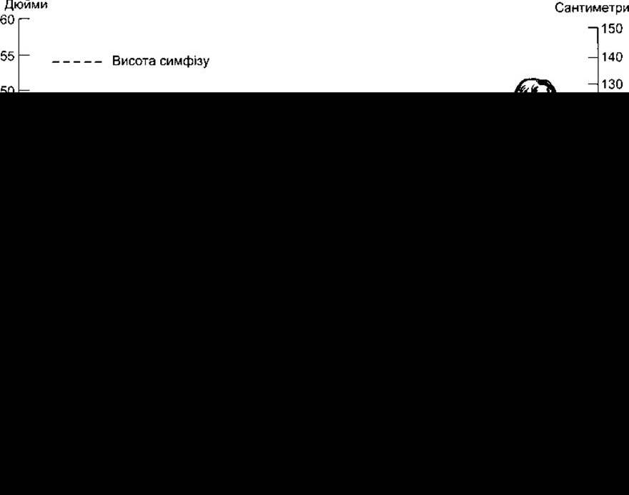
Fig. 22-15. Normal and pathological growth. Hypothyroid dwarfs (cretins) retain an infantile body Structure, whereas dwarfs of constitutional and, to a lesser extent, hypopituitary types have a body structure appropriate for their chronological age (reprinted with permission from Wilkins L: The Diagnosis and treatment of Endocrine Disorders in Childhood and Adolescence, 3rd ed/ Thomas, 1966).
The most common form of human dwarfism is Achondroplasia. It is characterized by shortened limbs with a normal trunk. This autosomal dominant disorder is caused by a mutation in the gene encoding fibroblast growth factor receptor 3 (FGFR3). FGFR3 belongs to the fibroblast growth factor receptor family and is normally expressed in cartilage tissue and the Brain.
Last update: 10/08/2026
Editorial and Educational Adaptation: This material has been compiled based on the primary/original source text. The project team performed an editorial review, corrected technical inaccuracies, structured sections, and adapted the content for an educational format.
What was processed:
- elimination of formatting defects (OCR errors, structural breaks, corrupted characters);
- editorial organization of content;
- standardization of terminology in accordance with academic sources;
- verification of factual statements against the original source text.
All mentions of the author, publication year, and origin of the primary text have been preserved in accordance with the source.