Orthopedics - Oleksa A.P. 2006
Congenital upper limb malformations
Congenital forearm malformations
Clubhand
Radial clubhand is a congenital upper extremity malformation caused by the underdevelopment of one of the paired forearm bones, or more rarely both, and may also be associated with Arthrogryposis, traumatic defects of the radius, etc.
Unilateral radial clubhand (Fig. III) resulting from radial hypoplasia is most common, while bilateral involvement is rarer. In bilateral cases, the radius is hypoplastic in one arm and completely absent in the other (Novachenko N.P., 1968).
Class="center">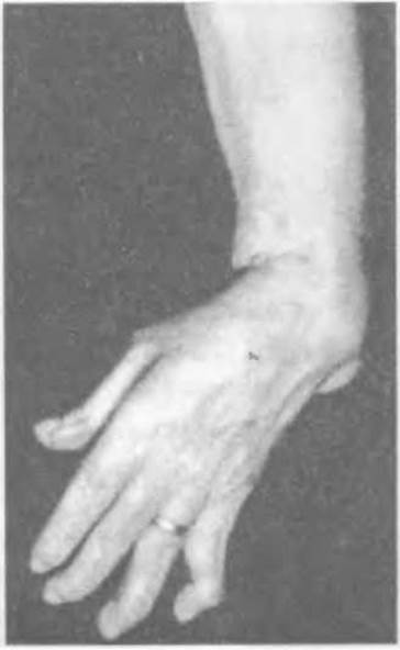
Fig. 111. Radial clubhand.
In most cases of congenital radial clubhand, the proximal part of the radius is reduced to varying lengths or represented only by a rudiment (Fig. 112). This condition leads to clubhand (manus valga congenita) because the ulna maintains nearly normal longitudinal growth, yet grows in an arched fashion since the normally developed hand shifts laterally in the absence of the radius as the primary support. Over time, subluxation or dislocation develops at the radiocarpal joint.
Very rarely, clubhand is accompanied by carpal bone hypoplasia, absence of the thumb along with its corresponding first metacarpal bone, or thumb hypoplasia (Zarichnyi V., 1977).
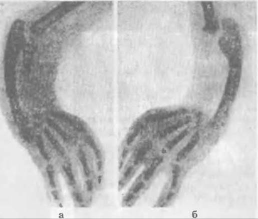
Fig. 112. Bilateral congenital clubhand: a - radial rudiment, b - absence of the radius (Novachenko M.P., 1968).
According to Dikhno A.M. (1940), ulnar clubhand (manus vara congenita) is observed seven times less frequently and is caused by ulnar hypoplasia or a defect of its distal part.
Under conditions of ulnar hypoplasia or defect, the radius grows and thickens, curving medially in an arc toward the underdeveloped ulna. As a result, the upper extremity becomes shortened.
As the child grows, this curvature progressively increases, causing the hand to deviate more and more toward the underdeveloped bone, sometimes forming an angle of 90° or greater relative to the forearm axis. Ulnar clubhand may also be combined with other congenital skeletal anomalies.
Clinically, diagnosing clubhand at any patient age presents no difficulty. However, radiography of the upper extremity bones is performed in all cases to determine the exact degree of underdevelopment of one forearm bone and curvature of the other, the status of the articulation with the Carpal Bones, and the presence of other hand developmental anomalies.
As a rule, all abnormalities detected radiographically are typical of clubhand, but radiological examination facilitates the Selection of the optimal Treatment method for patients.
Treatment. Once clubhand is diagnosed in early childhood, comprehensive conservative treatment is prescribed, aimed at preventing the rapid progression of the deformity. It is advisable to start treatment as early as possible, while the Tissues are pliable and before contractures have developed. Children undergo upper extremity Muscle massage, corrective Therapeutic Exercises for the hand and forearm, followed by immobilization in a plaster cast in the correct position after manipulations. Thermal Procedures (ozokerite-paraffin therapy, UHF, etc.) applied prior to massage and remedial gymnastics are beneficial. Such treatment is administered in courses with 2-3 week intervals, while night plaster or plastic splints
are used continuously. Mechanotherapy is performed daily.
As Novachenko M.P. (1968) points out, conservative treatment for children is carried out up to 10–12 years of age, and only thereafter is Surgical treatment performed. Since these operations are technically complex and involve bone work, they were not performed on young children in the past. With the advent of modern Anesthetic Management, the situation has changed.
The main objective of the various proposed surgeries is to realign the forearm axis and stabilize the hand in a functionally advantageous position.
To correct radial clubhand, Sayre and Parker proposed a two-stage operation. The First stage involves straightening the ulnar axis, while the second corrects THE POSITION OF the hand and fixes it in that position. This is achieved by performing an osteotomy of the curved ulna, followed by the resection of several carpal bones into whose bed the distal end of the ulna is transferred. However, because bony fusion between the ulna and carpal bones failed to occur, leading to recurrences of radial deviation of the hand, this Procedure did not gain widespread acceptance.
Lexer proposed creating a cleft in the carpal bones using an osteotome, including an osteotomy of the Base of the third metacarpal bone. Into this cleft, he inserted the sharpened end of the mobilized ulna to induce bony fusion. However, the latter did not occur because the necessary fixation devices were not available at the time.
Hohmann performed Lexer's operation, but supplemented it with an osteotomy and realignment of the axis of the arced-bent ulna.
Diagrams of the procedures by Romano and Hoff are presented in Figure 113. They consist of osteotomies of the distal PARTS OF THE ulna and their displacement, along with the hand, into a relatively functionally advantageous position. These operations are no longer used because they are difficult to perform in a way that places the hand in the desired position, cause significant forearm shortening, and yield unsatisfactory results.
Bardenheuer proposed using a resection knife to split the distal end of the ulna into two halves, displacing its radial part sideways and fixing it into a created groove in the scaphoid bone.
Antonelli proposed performing a similar longitudinal osteotomy of the ulna, but along its entire diaphysis, nearly to the elbow joint. After placing the hand in a normal position, the radial half was mobilized and wedged into the groove of the scaphoid bone. To prevent Displacement of the bone fragments, they were wired together. This interosseous space was filled with Muscles.
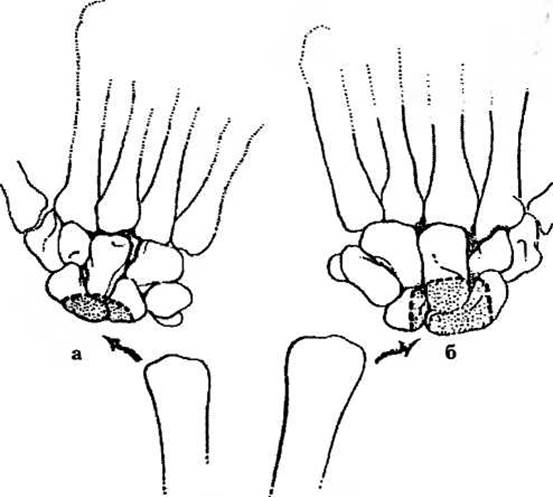
Fig. 113. Resection of carpal bones for transposition into the socket of the ulnar HEAD (a, b).
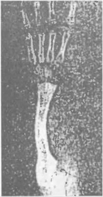
Fig. 114. Formation of the articular notch in the ulna on the radial side (Zarichnyi V., 1983).
Albee performed an osteoplastic procedure: after Setting the hand in the correct position and performing an osteotomy of the ulna in its middle third, he inserted a bone autograft ( harvested from the Tibia) between the osteotomy site and the scaphoid bone. To prevent displacement, the graft was seated into a depression in the scaphoid and secured with wire in the area of the ulnar osteotomy.
A. Kozlovsky proposed a surgical method similar to that of Albee. The difference is that Albee used a free bone graft, whereas Kozlovsky performed an oblique osteotomy of the ulna in its distal part and displaced the central fragment toward the scaphoid.
V. Zarichnyi shifts the ulna into the cavity formed after resecting the greater half of the scaphoid and removing the lunate; when necessary—specifically, if a pronounced arcuate curvature of the ulna is present—he aligns its axis following osteotomy and osteosynthesis.
Surgical technique. A transverse dorsal incision in the wrist joint area is used to expose the first row of carpal bones and the ulnar head.
In congenital clubhand resulting from the absence of the radius, the forearm muscles are underdeveloped, the soft tissues are tense, and the Joint Capsule is thickened. This creates certain difficulties when shifting the ulnar head into the cavity created after resecting the greater portion of the scaphoid and removing the lunate (Fig. 114).
To ensure better fusion of the ulna with the carpus and prevent displacement, the radial part of the scaphoid is preserved, and the ulna is fixed to the carpus using a thick Kirschner wire. Fixation of the ulna with wires is particularly important in children, as a plaster cast alone cannot prevent its displacement. Postoperatively, the arm is immobilized in a plaster cast with the forearm slightly flexed and pronated. The wires are removed after three weeks, and the plaster splint is discarded three months post-surgery. Rehabilitation therapy is then administered, and at night, the limb is placed in a corrective splint.
As the child grows, the ulna—transferred to the center of the wrist's load-bearing axis—remodels in accordance with Wolff's law, becoming thicker and resembling the distal end of the radius. If ulnar osteotomy and axial alignment are not performed, the arcuate curvature of the bone will progress during growth. Therefore, it is essential to restore the normal axis of the ulna in a timely manner.
All of the aforementioned procedures are applied in cases of congenital clubhand accompanied by the absence of the radius. If the proximal end of the radius is present, defect replacement using a bone graft is performed depending on its length and shape.
M.P. Novachenko successfully matched the suitably prepared proximal portion of the radius in three patients with a long bone autograft harvested from the Fibula, including its head and growth plate. After positioning the hand in a functionally favorable position, the fibular head was braced against the scaphoid and lunate bones, while the other end was connected to the rudimentary proximal end of the radius. Preserving the growth plate was intended to allow for continued longitudinal bone growth.
E.K. Nikiforova used an allograft with the fibular head during surgery based on the Albee method.
The question arises as to the optimal age for operating on children with congenital clubhand. M.P. Novachenko (1968) previously pointed out that surgical treatment for congenital clubhand can be performed in children older than 10–12 years.
As already mentioned, the deformity progresses as the child grows, creating The Need for surgery in early childhood.
With modern surgical techniques and anesthetic support, performing operations in early childhood is fully justified, as it prevents the progression of deformity during growth, The Development of fixed contractures and tissue tightening, and also facilitates the execution of surgeries while improving their anatomical and functional outcomes.
Beginning in 1963, V. Zarichnyi performed surgeries on a girl with bilateral clubhand and absent thumbs. The surgery on one hand was performed when the child was 10 months old, and on the other at 15 months of age. The distal ends of the ulnae were transferred into a cavity in the proximal carpal row after resecting 2/3 of the scaphoid and removing the lunate bones.
The distal end of the ulna gradually remodeled, becoming wider (almost like the radius) and capable of bearing weight. Due to the progression of the arcuate curvature of the ulnar diaphysis in the left arm (Fig. 115), corrective braces were used for an extended period, and at eight years of age, the girl underwent revision surgery. A ligament was reconstructed on the ulnar side by advancing a tendon autograft (from the extensor of the fifth toe) through a tunnel in the distal end of the ulna to the base of the fifth metacarpal bone. This improved the appearance of the hand and provided joint stability, although it slightly reduced the range of motion. However, because the curvature persisted, the patient underwent an osteotomy of the left ulna in its middle third at 12 years of age to align its axis. Long-term functional results of the surgeries are good.
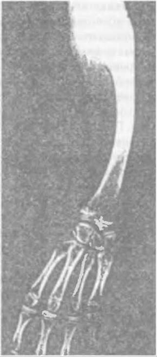
Fig. 115. Curvature of the ulna.
In addition to congenital clubhand, post-traumatic clubhand occurs as a result of radial defects. This can happen due to Osteomyelitis complications in open fractures, primary traumatic diaphyseal defects, and malunited fractures of the radius.
Untimely correction of post-traumatic clubhand leads to impaired hand function, weakness, and the development of deforming osteoarthritis of the radioulnar joint.
In cases of radial diaphyseal defects following osteomyelitis, resection within healthy bone limits with the removal of diseased fragment ends has proven effective. After the wound heals, the Ilizarov apparatus should be applied to lower the distal fragment via distraction until the radioulnar joint, forearm length, and proper hand position are fully restored. Then, without removing the apparatus, the defect is reconstructed using a bone autograft. The autograft must overlap the ends of both fragments, applied with its cancellous surface onto their corticated surfaces and secured tightly with cerclage wire. Currently, the method of bone defect reconstruction using an Ilizarov distraction regenerate is more frequently employed. To achieve this, without removing the apparatus, an osteotomy of the longer fragment of the radius is performed, and the defect is filled by translating the middle fragment until it docks with the shorter one. Bone union and remodeling of the distraction regenerate at the osteotomy site occur simultaneously. Restoring the length of the radius and filling the defect are crucial.
Similar procedures are performed for primary traumatic defects of the ulna.
In malunited fractures featuring angular fragment displacement and shortening of the radius, surgical tactics are chosen on an individual basis for each specific case. The primary goal is to restore the axis of the radius and its normal length.
As a rule, following osteotomy in the callus area, the radius remains somewhat shortened because manual reduction of the displacement is impossible. In such cases, the Ilizarov apparatus must be applied to restore the length of the radius via distraction and correct both the angular displacement and radial deviation of the hand. Once achieved, the apparatus is stabilized until bone union occurs. If a bone defect forms following distraction, a second elective surgery is performed to reconstruct the defect using either a bone graft or the Ilizarov technique. We have achieved good functional results with such procedures in six adult patients with post-traumatic clubhand.
To prevent hand deformity and functional impairment following fractures of the middle and lower thirds of the radius, the fragments must be perfectly repositioned using conservative or Surgical Methods, alongside the proper management of open fractures and their complications.
Last update: 10/08/2026
Editorial and Educational Adaptation: This material has been compiled based on the primary/original source text. The project team performed an editorial review, corrected technical inaccuracies, structured sections, and adapted the content for an educational format.
What was processed:
- elimination of formatting defects (OCR errors, structural breaks, corrupted characters);
- editorial organization of content;
- standardization of terminology in accordance with academic sources;
- verification of factual statements against the original source text.
All mentions of the author, publication year, and origin of the primary text have been preserved in accordance with the source.