Human Anatomy - Kotsan I. Ya. 2009
Skin
Skin Appendages
Hair and Nails are derivatives of the Skin.
Hair (pili) is a derivative of the epidermis. In humans, almost the entire skin surface (except for the red part of the Lips, palms, soles, and certain genital areas) is covered with hair. Hair is most developed on the scalp, eyebrows, pubis, and in men, also on the face, chest, and legs.
Hair is classified into the following types:
✵ long hair (scalp, mustache, beard);
✵ stiff hair (eyebrows, eyelashes);
✵ vellus hair (on most of the skin surface).
Each hair (Fig. 285) consists of a shaft (the free part protruding above the skin surface) and a ROOT (located within the thickness of the skin).
The length of the shaft ranges from 1–2 mm to 1.5 m, and its thickness ranges from 0.005 to 0.6 mm.
Class="center">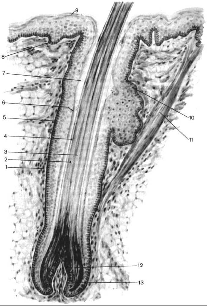
Fig. 285. Structure diagram of a hair (after V. G. Yeliseyev et al.)
1 — hair follicle; 2 — hair cortex; 3 — hair medulla; 4 — cuticle; 5 — outer root sheath; 6 — two layers of the inner root sheath; 7 — hair infundibulum; 8 — basal (germinal) layer of the epidermis; 9 — stratum corneum of the epidermis; 10 — sebaceous gland; 11 — arrector pili Muscle; 12 — hair bulb; 13 — hair papilla
The hair root is housed in a hair follicle, which is formed by an epithelial sheath and a Connective Tissue hair sac. The arrector pili muscle attaches to the sac. A sebaceous gland opens into the sac. The hair root extends into an expanded hair bulb, into which a connective tissue hair papilla invaginates. The papilla contains numerous capillaries that nourish the hair bulb. Situated above the papilla is the matrix, which is the growth zone of the hair. The hair grows through the division of Cells in the germinal zone, which then migrate upward.
The hair shaft consists of the cortex, medulla, and cuticle. The medulla is located in the central part of the hair and is completely absent in vellus hair. The cortex makes up the bulk of the hair mass and consists of elongated keratinized cells and pigment. The cuticle covers the hair externally and consists of anucleated keratinized scales arranged like shingles. Hair color depends on The amount of melanin pigment contained in the cortex, as well as on the air content within the hair and The properties of the cuticle itself. With age, the number of air bubbles increases while pigment synthesis ceases, resulting in graying hair.
Human hair grows continuously. It is estimated that a hair grows by 0.2–0.4 mm per day. The lifespan of a hair ranges from 2–3 months to 2–3 years. Fallen hairs are imperceptibly replaced by new ones. Under normal conditions, a healthy person loses 30–40 scalp hairs daily. Hair loss depends on a person's health status and the season (more hair is shed in autumn and spring).
Hair reduces skin heat loss and performs a protective function.
The nail (unguis) is a keratinized plate of the epidermis located on the dorsal surface of the distal Phalanges of the fingers and toes (Fig. 286). The nail bed (matrix unguis) is formed by the deep layers of the epidermis and the dermis.
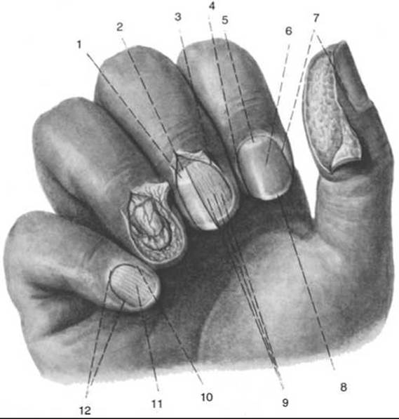
Fig. 286. Nails (right hand). The skin area with subcutaneous adipose tissue has been removed from the thumb (after R. D. Sinelnikov)
1 — nail root; 2 — hidden edge; 3 — nail bed groove; 4 — lateral edge; 5 — lunula; 6 — nail body; 7 — nail groove; 8 — free edge of the nail; 9 — nail bed ridges; 10 — nail bed furrow; 11 — nail bed; 12 — nail fold
A nail comprises the body (corpus unguis), the root (radix unguis), and four edges: the free edge (margo liber), which projects beyond the fingertip and can be trimmed; the hidden edge (margo occultus), located in the proximal region of the nail; and a paired lateral edge (margo lateralis). The skin cleft into which the hidden edge of the nail extends is called the nail sinus (sinus unguis). The lateral edge is surrounded by the nail fold (vallum unguis) and is partially positioned beneath this fold. The white strip of the nail body located near its hidden edge is called the lunula. The distal border of the lunula corresponds to the distal boundary of the germinal layer of the epidermis (stratum germinativum unguis), or hyponychium, where the multiplication and keratinization of the epidermal cells that form the nail take place. Nails grow continuously and quite rapidly, extending by 4 mm each month. The eponychium covers the nail root from above. The function of the nails is to protect the distal phalanges of the fingers and toes from mechanical injury.
Last update: 08/08/2026
Editorial and Educational Adaptation: This material has been compiled based on the primary/original source text. The project team performed an editorial review, corrected technical inaccuracies, structured sections, and adapted the content for an educational format.
What was processed:
- elimination of formatting defects (OCR errors, structural breaks, corrupted characters);
- editorial organization of content;
- standardization of terminology in accordance with academic sources;
- verification of factual statements against the original source text.
All mentions of the author, publication year, and origin of the primary text have been preserved in accordance with the source.