Human Anatomy - Kotsan I. Y. 2009
Central Nervous System. Spinal Cord
Meninges of the Spinal Cord
The Spinal Cord is externally enveloped by three membranes: the outer dura mater, the middle arachnoid mater, and the inner pia mater (Fig. 237).
The dura mater of the spinal cord (dura mater spinalis) is formed by dense Fibrous Connective Tissue. It is an elongated sac with fairly thick and robust walls, located within the vertebral canal and enclosing the spinal cord along with its anterior and posterior roots and other membranes. Superiorly, the spinal dura mater fuses with the margins of the foramen magnum and continues into the cranial dura mater, while its inferior part gradually tapers and terminates at the level of the II–III sacral vertebrae as the filum of the dura mater (external terminal filament), which attaches to the coccyx. Within the vertebral canal, the dura mater is anchored by extensions that continue into the perineural sheaths of the Spinal Nerves and fuse with the periosteum at each intervertebral foramen. Additionally, the spinal dura mater is reinforced by numerous fibrous bundles extending from the membrane to the posterior longitudinal ligament of THE Vertebral Column.
Class="center">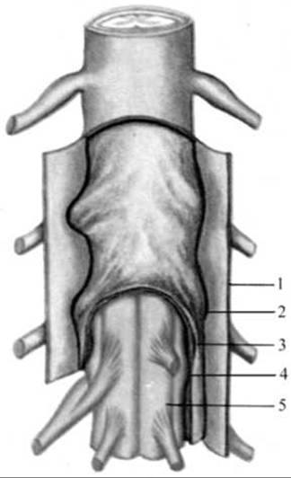
Fig. 237. Membranes of the spinal cord
1 — dura mater; 2 — subdural space; 3 — arachnoid mater; 4 — subarachnoid space; 5 — pia mater (vascular membrane) closely adhering to the spinal cord
Between the dura mater and the wall of the vertebral canal, which is formed by the vertebral periosteum and the ligaments of the vertebral column, lies the epidural space (spatium epidurale). The latter is filled with adipose tissue and contains the internal vertebral venous plexus, into which venous Blood drains from the spinal cord and vertebrae.
The arachnoid mater of the spinal cord (arachnoid mater spinalis) lines the inner surface of the dura mater and fuses with it near the intervertebral foramina. It is an extremely thin, translucent membrane constructed of Cytology/practical/45.html">Dense connective tissue, devoid of any Blood Vessels, and nourished by the CEREBROSPINAL FLUID contained within the subarachnoid space. The arachnoid mater is separated from the inner surface of the dura mater by the subdural space (spatium subdurale), which is traversed by numerous delicate connective tissue trabeculae. Superiorly, the subdural space of the spinal cord communicates freely with the corresponding space in the cranial cavity. Inferiorly, this space ends blindly at the level of the second sacral vertebra. The subarachnoid space (spatium subarachnoidale), filled with cerebrospinal fluid, separates the arachnoid mater from the pia mater. In the lower part of සුthe vertebral canal, the roots of the spinal nerves float freely within the subarachnoid space. In this region (below the second lumbar vertebra), it is safest to perform a needle puncture (without the risk of damaging the spinal cord) to collect cerebrospinal fluid for diagnostic purposes, the total volume of which is 120–140 ml. In the upper sections, the subarachnoid space of the spinal cord is continuous with the corresponding cranial subarachnoid space.
The pia mater of the spinal cord (pia mater spinalis) closely invests the spinal cord. It comprises two layers—inner and outer—between which blood vessels are situated. The inner layer fuses with the spinal cord tissue. Connective tissue fibers branching off from this membrane accompany blood vessels and penetrate with them into the substance of the Brain. The outer surface of the pia mater is covered by endothelial Cells that produce cerebrospinal fluid into the subarachnoid space. The subarachnoid space contains numerous bundles and lamellae connecting the arachnoid mater to the pia mater and the spinal cord. Extending from the lateral surfaces of the pia mater, between the anterior and posterior roots, right and left toward the arachnoid mater, is a thin, strong lamina known as the denticulate ligament (ligamentum denticulatum). This ligament has a continuous origin from the pia mater and laterally divides into 20–30 denticulations that fuse not only with the arachnoid mater but also with the dura mater of the spinal cord. The upper denticulation of this ligament is located at the level of the foramen magnum, and the lower one lies between the roots of the XII thoracic and 1st lumbar spinal nerves. Through this frontally positioned denticulate ligament, the spinal cord appears suspended within the cerebrospinal fluid-filled subarachnoid space. On the posterior surface of the spinal cord, along the posterior median sulcus, a sagittally oriented posterior septum (septum posterior) extends from the pia mater to the arachnoid. In the lumbar and sacral regions, where the cauda equina is located, the denticulate ligament and the posterior subarachnoid septum are absent. The adipose tissue and venous plexuses of the epidural space, the spinal Meninges, the cerebrospinal fluid, and the ligamentous apparatus significantly dampen The impact of various shocks and jolts experienced by the body during movement, protect it against mechanical injury, and participate in the fixation of the spinal cord.
Last update: 08/08/2026
Editorial and Educational Adaptation: This material has been compiled based on the primary/original source text. The project team performed an editorial review, corrected technical inaccuracies, structured sections, and adapted the content for an educational format.
What was processed:
- elimination of formatting defects (OCR errors, structural breaks, corrupted characters);
- editorial organization of content;
- standardization of terminology in accordance with academic sources;
- verification of factual statements against the original source text.
All mentions of the author, publication year, and origin of the primary text have been preserved in accordance with the source.