Human Anatomy, Part 1 - K. A. Dyubenko, A. K. Kolomiytsev, Y. B. Chaykovsky 2002
Special Section
Respiratory system, systema respiratorium [respiratory apparatus, apparatus respiratorius] – General Overview
Muscles that tense the vocal folds
1. The vocal Muscle, musculus vocalis, is a paired muscle that originates from the internal surface of the thyroid Cartilage and attaches to the vocal process of the arytenoid cartilage. The medial margin of the muscle is fused with the vocal ligament, while the lateral margin adjoins the thyroarytenoid muscle. Upon contraction, it pulls the vocal process forward. As a result, the vocal folds relax, and the rima glottidis narrows slightly.
2. The cricothyroid muscle, musculus cricothyroideus, is a paired muscle originating from the middle of the arch of the cricoid cartilage and inserting into the inferior margin (straight part, pars recta) and the inferior horn (oblique part, pars obliqua) of the thyroid cartilage. When contracted, it pulls the thyroid cartilage forward, which tensions the vocal folds.
Стінка гортані утворена її хрящами (об’єднаними за допомогою зв’язок, суглобів, м’язів), фіброзно-еластичною мембраною, слизовою оболонкою, зовнішньою сполучнотканинною оболонкою.
The fibro-elastic membrane of the Larynx, membrana fibroelastica laryngis, lies beneath the laryngeal mucosa. At the level between the upper margin of the cricoid cartilage arch, the inferior notch of the thyroid cartilage, and the vocal processes of the arytenoid cartilages, this membrane tapers from bottom to top, forming the elastic cone, conus elasticus.
The mucous membrane, tunica mucosa, is lined with pseudostratified ciliated columnar epithelium, except for the vocal folds, which are covered with non-keratinized stratified squamous epithelium. The epiglottis is also lined with non-keratinized stratified squamous epithelium because here the laryngeal mucosa is continuous with the mucosa of the digestive tract. The submucosa contains laryngeal glands and lymphoid nodules.
The external Connective Tissue coat covers the laryngeal cartilages, contains numerous elastic fibers, and essentially forms a part of the visceral layer of the Cervical fascia.
The laryngeal cavity, cavitas laryngis, forms a peculiar tube, widened superiorly and inferiorly, and narrowed in the middle, thus resembling an hourglass (Fig. 285). The cavity begins with the laryngeal inlet, aditus laryngis, which is bounded anteriorly by the epiglottis, laterally by the aryepiglottic folds, plicae aryepiglotticae, and posteriorly by the apices of the arytenoid cartilages.
The upper widened part of the laryngeal cavity is called the vestibule, vestibulum laryngis. It extends from the laryngeal inlet to the vestibular folds, plicae vestibuläres, which bound the rima vestibuli (Fig. 286). The mucous membrane of the laryngeal vestibule is highly sensitive—its irritation reflexively triggers a strong cough.
The middle narrowed part of the larynx extends from the vestibular cleft to the rima glottidis, formed by the vocal folds, plicae vocales. The rima glottidis is the narrowest part of the larynx. It consists of two parts. The anterior part, bounded by the sections of the vocal folds containing the vocal Ligaments and Muscles, is called the intermembranous part, pars intermembranacea. The posterior part, bounded by the sections of the vocal folds enclosing the vocal processes of the arytenoid cartilages, is called the intercartilaginous part, pars intercartilaginea. A recess, the laryngeal ventricle, ventriculus laryngis, is formed between the vestibular and vocal folds.
Class="center">
Fig. 285. Laryngeal cavity, cavitas laryngis: A - frontal section; Б - sagittal section.
The lower widened part of the larynx is the infraglottic cavity, cavitas infraglottica. Narrowing inferiorly, it continues into the Trachea. During laryngeal examination (laryngoscopy) in a living person, one can observe the vestibular and vocal folds, assess the condition of the mucous membrane, and evaluate the width of the rima glottidis. The mucosa appears smooth, has a uniform pink color, and exhibits a reddish hue in the region of the vestibular folds.
During quiet breathing, the rima glottidis is quite wide, whereas during sound production (phonation) it periodically narrows and may even close completely. Sound production occurs because active vibrations of the vocal folds during expiration generate oscillating air waves. The sound produced in the larynx is amplified and acquires its characteristic timbre in the resonators—the Upper Respiratory Tract, Oral Cavity, and Paranasal Sinuses.
Age-related Features of the larynx. In newborns, the larynx is short and wide, located three vertebrae higher than in adults. The corniculate cartilages are absent, and the laryngeal inlet is wide. The thyrohyoid ligaments are also absent. In subsequent years, the larynx increases in size and gradually descends. By age 7, all its anatomical structures are well-defined. By age 13, it reaches the level of the IV–VI cervical vertebrae. At 12–15 years of age, the larynx grows particularly intensively in boys, which leads to voice mutation. In girls, laryngeal growth occurs more gradually.
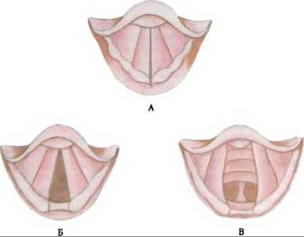
Fig. 286. Position of the vocal folds under various functional states:
А - rima glottidis closed; Б - rima glottidis open; В - rima glottidis widened
X-ray Anatomy of the larynx. In the lateral projection, the anterior and posterior contours of the walls of the larynx and Pharynx, the superior and posterior contours of the cricoid cartilage, the shadows of the vestibular and vocal folds, the laryngeal ventricles, and the epiglottis are visible.
In the sagittal projection, the lateral walls of the larynx, the shadows of the epiglottis, aryepiglottic folds, vestibular and vocal folds, and laryngeal ventricles can be observed.
Blood supply to the larynx is provided by the superior and inferior laryngeal Arteries, which are Branches of the corresponding thyroid arteries. Veins form plexuses within the mucous membrane and drain into the inferior and upper laryngeal veins, which in turn empty into the inferior and superior thyroid veins.
Lymphatic vessels of the larynx drain Lymph into the deep cervical Lymph Nodes.
Innervation of the larynx is provided by the inferior and superior laryngeal nerves (from the vagus nerves) and branches of the superior cervical ganglion of the Sympathetic trunk.
Last update: 08/08/2026
Editorial and Educational Adaptation: This material has been compiled based on the primary/original source text. The project team performed an editorial review, corrected technical inaccuracies, structured sections, and adapted the content for an educational format.
What was processed:
- elimination of formatting defects (OCR errors, structural breaks, corrupted characters);
- editorial organization of content;
- standardization of terminology in accordance with academic sources;
- verification of factual statements against the original source text.
All mentions of the author, publication year, and origin of the primary text have been preserved in accordance with the source.