Human Anatomy Part 1 - K. A. Dyubenko, A. K. Kolomiysev, Yu. B. Chaykovsky 2002
Special Section
Myology, myologia [the study of muscles] - General Overview
Myology, myologia (from Latin musculus – Muscle, and logos – study) is a branch of anatomy that studies the development, Structure, function, and topographic-Anatomical Features of Muscles.
Muscles, musculi, are anatomical structures composed primarily of muscular tissue. They enable the movement of the body and its parts in space, maintaining the upright posture characteristic of humans, as only muscles keep certain bones immobile relative to others in an upright body position. Muscles account for 42% of an adult's body weight, and over 20% in newborns and children. In individuals who train their musculature, this mass can reach up to 50% of the total body weight. The Human Body contains over 400 skeletal muscles (Fig. 113) that form The Muscular System, systema musculare.
Muscles are divided into striated (skeletal) and smooth. Striated muscles are involved in moving the body and skeletal parts, maintaining balance, posture, and joint stabilization. They are located around natural orifices and act as closing muscles [sphincters]. In the Pharynx and Esophagus, they ensure swallowing and food passage, while in the Larynx, they are responsible for sound production. The Heart muscle is classified separately among striated muscles. Smooth muscles are a structural component of the walls of Blood Vessels, Skin, and Internal Organs such as The Stomach, small and large intestines, and Uterus. The contraction of smooth muscles drives gastrointestinal motility, creating optimal conditions for Digestion and maintaining blood pressure at a certain level. In some internal organs and PARTS OF THE Circulatory system, smooth muscles function as closing muscles [sphincters].
Based on their shape, muscles are classified as long, broad, flat, short, and circular (Fig. 114 D–E). According to the arrangement of muscle fascicles relative to tendons, they are divided into unipennate, bipennate, and multipennate (Fig. 114, 1–4). Short muscles have equal length and width and are located between the vertebrae. Long muscles are predominantly fusiform and found in the upper and lower limbs. Long muscles feature tendons at their ends, through which they attach to bones and tightly fuse with the periosteum. Each muscle has an origin, origo, and an insertion point, insertio. Tendons vary in shape; for instance, in broad flat muscles, they form tendon expansions known as aponeuroses, aponeurosis.
Class="center">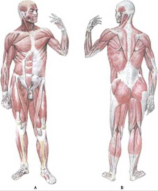
Fig. 113. Muscles of the human body:
A – anterior view; B – posterior view
Muscles are located on the bones of various skeletal regions and are accordingly classified by body region: Muscles of the Trunk (back, chest, and abdomen), HEAD and Neck, upper limb (shoulder girdle, arm, forearm, and hand), and lower limb (pelvis, thigh, leg, and FOOT). Muscles may cross one or several joints. Depending on this, they are distinguished as monoarticular, biarticular, and multiarticular muscles.

Fig. 114. Muscle shapes and the orientation of muscle fascicles:
1 – unipennate muscle; 2 – bipennate muscle; 3 – biceps muscle; 4 – triceps muscle; 5 – digastric muscle; 6 – circular muscle; 7 – rectus muscle; 8 – transverse muscle
Based on their functional characteristics, muscles are divided into synergists, which perform coordinated movements, and antagonists, which perform opposite actions.
Synergists include all muscles that act simultaneously on a joint during contraction while lying on the same side of its axis. Examples include the flexors of the forearm and arm, which perform flexion at the elbow joint. Antagonists include all muscles that function in the direction opposite to another muscle group. For instance, the flexors of the arm are antagonists to the extensors of the arm. The Functions of synergistic and antagonistic muscles can alternate. When performing flexion and extension in the wrist joint, the radial and ulnar flexors act on one side, while the extensors of the hand act on the other as antagonists. Conversely, when performing adduction and abduction of the hand, they become synergists.
Muscles are also characterized by indicators such as Anatomical and physiological cross-sectional area. The anatomical cross-sectional area is the surface area corresponding to a perpendicular cut across the length of the muscle. It passes through the muscle belly at its widest part and reflects the size and thickness of the muscle. The physiological cross-sectional area represents the total cross-sectional area of all muscle fibers that make up the muscle. The physiological cross-sectional area determines the strength of the muscle. According to P. F. Lesgaft, the absolute strength of a muscle is greater the larger the number of its muscle fibers; therefore, it is determined by the total number of muscle fascicles, or the so-called physiological cross-section.1
Based on The Nature of their primary movements, muscles are classified into: flexors, mm. flexores, and extensors, mm. extensores; adductors, mm. adductores, and abductors, mm. abductores; levators, mm. levatores, and depressors, mm. depressores; pronators, mm. pronatores, and supinators, mm. supinatores; constrictors, mm. constrictores, sphincters, mm. sphinctores, and dilators, mm. dilatatores; rotators, mm. rotatores; tensors, mm. tensores, and erectors, mm. erectores. Masticatory, facial expression (mimetic), and Respiratory Muscles are also distinguished.
Last update: 08/08/2026
Editorial and Educational Adaptation: This material has been compiled based on the primary/original source text. The project team performed an editorial review, corrected technical inaccuracies, structured sections, and adapted the content for an educational format.
What was processed:
- elimination of formatting defects (OCR errors, structural breaks, corrupted characters);
- editorial organization of content;
- standardization of terminology in accordance with academic sources;
- verification of factual statements against the original source text.
All mentions of the author, publication year, and origin of the primary text have been preserved in accordance with the source.