Human Anatomy Part 1 - K. A. Dyubenko, A. K. Kolomiytsev, Yu. B. Chaykovsky 2002
Special Section
Joints of the upper limb, juncturae membri superioris - Joints of the free upper limb, articulationes membri superioris liberi
The shoulder joint, articulatio humeri [glenohumeralis], connects the Pectoral Girdle to the free upper limb (Fig. 88). It is formed by the articular surface of the HEAD of the humerus, covered with hyaline Cartilage, and the glenoid cavity of the scapula, cavitas glenoidalis, which is three times smaller than the humeral articular surface; this anatomical feature provides a wide range of motion while predisposing the joint to frequent dislocations (F. V. Sudzylovsky).
There is a disparity between the articulating bone surfaces; therefore, to enhance congruence, a glenoid labrum, labrum glenoidale, forms along the rim of the glenoid cavity. The articular capsule is thin and spacious. It originates from the edge of the glenoid labrum and attaches to the anatomical neck of the humerus, leaving both tubercles of the bone outside the articular cavity.
The shoulder joint is the only joint whose cavity is traversed by the tendon of the long head of the biceps brachii Muscle. It lies within the intertubercular sulcus of the humerus and is surrounded by a synovial sheath. The joint is reinforced by the coracohumeral ligament, lig. coracohumerale, which arises from the coracoid process of the scapula and blends into the Joint Capsule.
The shoulder joint is bounded by Muscles on three sides: superiorly by the supraspinatus, posteriorly by the infraspinatus, teres major, and teres minor, anteriorly by the subscapularis, and laterally by the deltoid. The surrounding muscle tendons not only stabilize the joint during movement but also retract its capsule.
According to the shape of its articular surfaces, the shoulder joint is classified as a ball-and-socket (spheroidal) joint. Movements are possible around three mutually perpendicular axes: the sagittal axis (abduction and adduction), the vertical axis (pronation and supination), and the frontal axis (flexion and extension). Circumduction, circumductio, and a complex movement known as elevation (raising the arm upward) are also possible.
Class="center">
Fig. 88. Shoulder joint, articulatio humeri: A - posterior surface; B - frontal section
Due to extensive mobility and relatively weak fixation, the shoulder joint is prone to dislocations (complete Displacement of the articular bone surfaces beyond the physiological norm).
Blood supply is provided by the articular network, rete articulare, formed by Branches of the anterior circumflex humeral artery, a. circumflexa humeri anterior, posterior circumflex humeral artery, a. circumflexa humeri posterior, and thoracoacromial artery, a. thoracoacromialis (from the axillary artery, a. axillaris). Venous drainage flows into corresponding Veins that empty into the axillary vein, v. axillaris.
Lymphatic drainage occurs via deep Lymphatic vessels into the axillary Lymph Nodes, nodi lymphatici axillares.
Innervation of the joint capsule is supplied by the axillary nerve (Note: original text mentions v. axillaris, kept as per rule 3/context if referring to vascular/neural bundles, but strictly translating the literal term).
Radiographic anatomy. Radiographs of the shoulder joint in the anteroposterior projection clearly reveal the bones participating in its formation (Fig. 89). The greater and lesser tubercles of the humerus are distinct. The shadow of the coracoid process appears rounded and projects onto the scapular axis. A clear space is visible between the humeral head and the glenoid cavity of the scapula, cavitas glenoidalis scapulae. In children, epiphyseal lines are visible at the proximal end of the humerus.
The elbow joint, art. cubiti [cubitalis], is a compound joint, art. composita, formed by three articulations: the humeroulnar, humeroradial, and proximal radioulnar joints. They all share a common cavity and are enclosed by a single joint capsule (Fig. 90).
The humeroulnar joint, art. humero-ulnaris, is a hinge joint, ginglymus, formed by the trochlea of the humerus, trochlea humeri, and the trochlear notch of the ulna. The articular surfaces feature a helical (screw-like) inclination.
The humeroradial joint, art. humeroradialis, is formed by the capitulum of the humerus, capitulum humeri, and the articular fovea on the head of the radius. It is a ball-and-socket (spheroidal) joint, art. spheroidea.
The proximal radioulnar joint, art. radioulnaris proximalis, is formed by the radial notch of the ulna, incisura radialis, and the articular circumference of the radius, circumferentia articularis radii. It is a cylindrical joint. Movements in the elbow joint occur around two mutually perpendicular axes: the frontal axis (flexion and extension) and the vertical axis, which passes through the humeroulnar joint (pronation and supination).
The elbow joint is reinforced by the following ligaments:
- annular ligament of the radius, lig. anulare radii, which forms a ring encircling the head of the radius and holding it firmly within the radial notch of the ulna;
- radial collateral ligament, lig. collaterale radiale, extending from the lateral epicondyle of the humerus to the head of the radius;
- ulnar collateral ligament, lig. collaterale ulnare, extending from the medial epicondyle of the humerus to the margin of the trochlear notch of the ulna.
On the anterior aspect of the humerus, the joint capsule lies 1.5–2 cm above the coronoid and radial fossae; posteriorly, it lies above the olecranon fossa, encompassing its sides, and then runs along the margins of the medial and lateral epicondyles, leaving them free. Inferiorly, it attaches to the neck of the radius and the margin of the trochlear notch of the ulna. The joint capsule of the elbow is thin both anteriorly and posteriorly.
Blood supply is derived from the articular network of the elbow, rete articulare cubiti, formed by the superior and inferior ulnar collateral Arteries (from the brachial artery, a. brachialis), middle collateral and radial collateral arteries (from the profunda brachii artery, a. profunda brachii), radial recurrent artery (from the radial artery, a. radialis), posterior recurrent interosseous artery (from the posterior interosseous artery, a. interossea posterior), and anterior and posterior ulnar recurrent arteries (from the ulnar artery, a. ulnaris). Venous drainage is carried out by companion veins into the deep VEINS OF THE upper limb—the radial, ulnar, and brachial veins (vv. radiales, ulnares, brachiales).
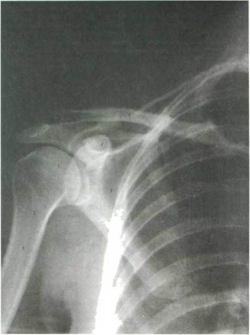
Fig. 89. Overview radiograph of the shoulder joint (32-year-old female) - anteroposterior projection (after Ya. I. Kryzhanovsky)
1 - clavicula, 2 - acromion, 3 - scapula; 4 - processus coracoideus; 5 - caput humeri; 6 - tuberculum majus, 7 - collum chirurgicum; 8 - os humerus

Fig. 90. Elbow joint, articulatio cubiti: A - anterior view; B - medial view
Lymph drainage is carried out via deep lymphatic vessels into the cubital lymph nodes (nodi lymphatici cubitales) and axillary lymph nodes (nodi lymphatici axillares).
Innervation is provided by branches of the median nerve (n. medianus), radial nerve (n. radialis), ulnar nerve (n. ulnaris), and musculocutaneous nerve (n. musculocutaneus).
X-ray anatomy. On an anteroposterior (AP) radiograph of the elbow joint, the humeroulnar and humeroradial joint spaces, the lateral and medial epicondyles, and the trochlea and capitulum of the humerus are clearly visible (Fig. 91). The olecranon and coronoid fossae overlap each other. In the lateral view, the humeroulnar joint space is visible along its entire length. Cartilaginous epiphyseal plates persist in the elbow joint until the age of 15–17.
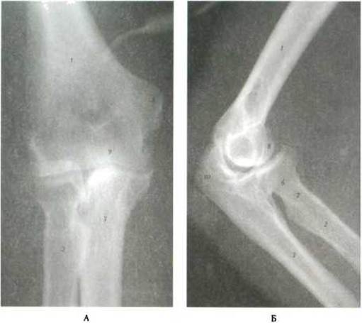
Fig. 91. Plain radiograph of the right elbow joint (in a 45-year-old male) (after Ya. I. Kryzhanovsky): A - anteroposterior projection; B - lateral projection
1 - os humerus; 2 - radius, 3 - ulna; 4 - epicondylus medialis; 5 - epicondylus lateralis; 6 - caput radii; 7 - collum radii; 8 - capitulum humeri; 9 - trochlea humeri; 10 - olecranon
Last update: 08/08/2026
Editorial and Educational Adaptation: This material has been compiled based on the primary/original source text. The project team performed an editorial review, corrected technical inaccuracies, structured sections, and adapted the content for an educational format.
What was processed:
- elimination of formatting defects (OCR errors, structural breaks, corrupted characters);
- editorial organization of content;
- standardization of terminology in accordance with academic sources;
- verification of factual statements against the original source text.
All mentions of the author, publication year, and origin of the primary text have been preserved in accordance with the source.