Nephrology for the Family Physician - O.I. Bakaliuk 2003
Emergency Uro-Nephrology
Testicular and Spermatic Cord Torsion
This condition occurs most frequently at ages 10-15 and 60-70 years. It is associated with predisposing factors such as testicular hypermobility (cryptorchidism, delayed testicular descent and ectopia, congenital elongation of the Spermatic Cord, Separation of the Epididymis and Testis, etc.) or specific circumstances (rapid growth in children, wearing tight clothing, sudden herniation of abdominal viscera in inguinoscrotal hernias), which may cause the testis to twist around its vertical or horizontal axes. Such torsion can be triggered by scrotal trauma, sudden movement, abrupt straining of the Abdominal Muscles, or forceful coughing. The primary risk factor for testicular torsion is a congenital anomaly known as the "bell-clapper deformity" (G. Tamkin, 2000).
Torsion of the spermatic cord and testis rapidly impairs Blood AND Lymph Circulation, leading to total hemorrhagic necrosis. Irreversible testicular changes in children develop within just 6-8 hours from the onset of symptoms, which necessitates both timely Diagnosis and emergency medical intervention.
The Clinical presentation of testicular torsion is typical, characterized by the sudden onset of acute testicular pain (triggered by the aforementioned factors) radiating to the inguinal region. The pain may be accompanied by Acute Urinary Retention, nausea, vomiting, fever, and circulatory collapse. Scrotal Asymmetry with Skin erythema develops rapidly. The testis is drawn upward toward the ROOT of the Scrotum, becomes enlarged, and assumes a horizontal lie. The spermatic cord is also thickened; Palpation of both the cord and the testis is exquisitely painful, and the cremasteric reflex is absent. Intra-abdominal positioning of the testis presents with symptoms of an "acute abdomen" without local scrotal findings, whereas inguinal cryptorchidism manifests as Swelling and tenderness of the soft Tissues in the Inguinal Canal region. In some cases, the testis can be "transilluminated" through the skin as a dark-red mass, and its palpation remains extremely painful. Differential diagnostic features distinguishing orchitis from testicular torsion include Prehn's sign (J. Prehn): in orchitis, elevating the scrotum relieves the pain, whereas in testicular torsion it does not; to differentiate between testicular torsion and epididymitis, Roce's sign is used (A.E. Roce: in testicular torsion, the epididymis is not palpable, whereas in epididymitis, the boundary between the enlarged epididymis and the testis can be clearly delineated).
In addition to clinical findings, diagnostic evaluation of testicular torsion involves Doppler Ultrasonography (absence of pulsatile blood flow signals on the affected side, unlike inflammatory conditions where perfusion on the affected side increases) and dynamic Tc99 scintigraphy (marked reduction in radiotracer uptake on the affected side). Diagnosis of an undescended intra-abdominal testis also takes into account its absence within the scrotum.
Table 12 presents the differential diagnostic criteria for various pathological conditions accompanied by scrotal enlargement and pain (H.L. May, 1984; G. Tamkin, 2000).
Class="center">Clinical characteristics of pathological conditions associated with scrotal enlargement and pain (after H.L. May, 1984)
|
Condition |
Age |
Pain characteristics |
Onset |
|
|
1 |
2 |
3 |
4 |
5 |
|
Hydrocele |
Pediatric |
Absent |
Scrotal contents transilluminate through the skin. An acute, painful hydrocele in young adults may be associated with trauma, inflammation, or testicular tumor |
|
|
Testicular tumor |
25-30 yrs. |
Absent |
Contents do not transilluminate through the skin. Gynecomastia may be observed in 10% of cases. Acute pain occurs in the event of testicular infarction |
|
|
Testicular torsion |
< 25 yrs. |
Severe |
Sudden |
Anterior epididymitis is observed. Cremasteric reflex (+). Urgent care required |
|
Epididymitis |
> 20 yrs. |
Severe |
Gradual |
Posterior epididymitis is observed; typically pyuria, episodes of prostatitis. Cremasteric reflex (+). Most common causes include instrumentation, strictures, Urinary Tract infection, recent prostatectomy |
|
Orchitis |
> 20 yrs. |
Moderate to severe |
Variable, usually gradual |
Usually preceded by mumps |
|
> 20 yrs. |
Absent or moderate |
Gradual |
Exacerbated in the upright position. Acute onset indicates acute venous obstruction |
|
|
Strangulated scrotal hernia |
Elderly |
Moderate to severe |
Sudden |
Bowel sounds may occasionally be auscultated over the scrotum. Urgent care required |
The only way to save a torsed testis from necrosis is untwisting (detorsion) of the spermatic cord using two approaches: conservative and surgical.
Conservative testicular detorsion is performed as follows (Ya.B. Yudin et al., 1980). The patient lies in a supine position. Detorsion is carried out in the direction opposite to the twist—counterclockwise for the right testis, and clockwise for the left testis.
The median raphe of the scrotum serves as a landmark for determining the direction of detorsion. The testis, along with the scrotal tissues, is grasped and rotated 180° away from the median raphe of the scrotum (Fig. 108).
Simultaneously, gentle downward traction is applied to the testis.
The testis is then released, and the maneuver is repeated several times. Successful detorsion is indicated by a reduction in scrotal pain, restoration of testicular mobility, and its descent back into its normal anatomical position.
If conservative detorsion fails (within 2-5 minutes), the patient must be taken to surgery immediately (surgical detorsion with orchiectomy in cases of testicular necrosis).
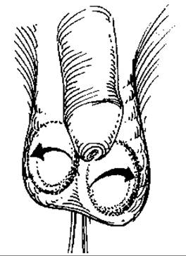
Fig. 108. Conservative testicular detorsion.
Last update: 08/08/2026
Editorial and Educational Adaptation: This material has been compiled based on the primary/original source text. The project team performed an editorial review, corrected technical inaccuracies, structured sections, and adapted the content for an educational format.
What was processed:
- elimination of formatting defects (OCR errors, structural breaks, corrupted characters);
- editorial organization of content;
- standardization of terminology in accordance with academic sources;
- verification of factual statements against the original source text.
All mentions of the author, publication year, and origin of the primary text have been preserved in accordance with the source.