BIOCHEMISTRY - L. Stryer - 1984
VOLUME 2
PART II GENERATION AND STORAGE OF METABOLIC ENERGY
CHAPTER 19. PHOTOSYNTHESIS
19.21. Halobacterial Purple Membrane Protein Pumps Protons for ATP Synthesis
Valuable insights into The Mechanism of Photophosphorylation and Oxidative Phosphorylation have come from studies of photosynthetic processes in halobacteria, which possess a light-absorbing group distinct from chlorophyll. These Bacteria require high concentrations of NaCl for growth, with an optimum of 4.3 M (ordinary seawater contains 0.6 M NaCl). Halobacteria are found in natural salt lakes and in evaporation ponds used to produce salt from seawater, such as those in San Francisco Bay near the airport. These bacteria have a Cell envelope consisting of a membrane surrounded by a glycoprotein Cell wall. Walther Stoeckenius fractionated this cell membrane into yellow, red, and purple fractions. The yellow fraction consisted mainly of the walls of gas vacuoles, which enable the bacteria to reach their preferred depth in the Water Column. The red fraction contains the Respiratory Chain and other enzyme systems for oxidative phosphorylation, as well as a protective red pigment that shields the bacteria from the lethal effects of blue light. The purple fraction consists of the purple membrane protein (26 kDa) and forty associated Lipids. This protein is also called Bacteriorhodopsin because, like rhodopsin—the visual pigment of vertebrate retinas (Section 37.13)—it contains retinal as a chromophore. The retinal group is attached to a Lysine side chain of the protein via a protonated Schiff base linkage.
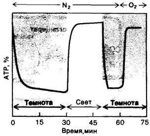
In the presence of O2, halobacteria synthesize ATP via oxidative phosphorylation. Under oxygen-depleted conditions, they switch to a photosynthetic mechanism (Fig. 19.26). Stoeckenius and his coworkers demonstrated that The Role of the purple membrane protein is to pump protons from the inside to the outside of The Cell upon illumination. Indeed, the purple membrane protein spans The cell membrane (Fig. 10.32 in Section 10.18), as would be expected for a proton pump. Flame spectroscopy studies have shown that light converts the retinal Schiff base linkage from a protonated form with an absorption band at 560 nm to a deprotonated form with a peak at 412 nm. This reaction cycle takes only a few milliseconds, and under intense illumination, each molecule of the purple membrane protein can pump several hundred protons per second. The proton-motive force generated in this manner is subsequently utilized by the ATP-synthesizing assembly (F0 F1) to phosphorylate ADP within a spatially distinct region of the red membrane. The F0-F1 complex is oriented such that its globular F1 component faces the cell interior. The exact same ATP-synthesizing assembly is utilized in both OXIDATIVE PHOSPHORYLATION AND photophosphorylation. The proton-motive force generated by either process can also drive The Active Transport of ions and Amino Acids into halobacteria.
Class="center">Fig. 19.26. Experiment demonstrating that illuminated halobacteria can synthesize ATP in the absence of O2
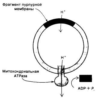
The availability of a purified purple membrane fraction enabled Stoeckenius and Efraim Racker to carry out a highly informative reconstitution experiment. They produced synthetic phospholipid vesicles containing the purple membrane protein from halobacteria and the ATP-synthesizing F1-F0 complex from bovine Heart Mitochondria (Fig. 19.27). Illumination of these vesicles resulted in The formation of a transmembrane proton gradient and the generation of ATP from ADP and Pi. ATP synthesis ceased upon The addition of proton carriers that disrupted the proton gradient, highlighting the essential role of the proton-motive force in this process. These reconstituted vesicles, containing only a proton pump and ATP synthase within a closed membrane impermeable to protons, serve as the simplest model system for chemiosmotic energy Transduction.
Fig. 19.27. Reconstituted membrane vesicles containing the purple membrane protein and the mitochondrial ATP-synthesizing complex generate ATP upon illumination
Last update: 06/08/2026
Editorial and Educational Adaptation: This material has been compiled based on the primary/original source text. The project team performed an editorial review, corrected technical inaccuracies, structured sections, and adapted the content for an educational format.
What was processed:
- elimination of formatting defects (OCR errors, structural breaks, corrupted characters);
- editorial organization of content;
- standardization of terminology in accordance with academic sources;
- verification of factual statements against the original source text.
All mentions of the author, publication year, and origin of the primary text have been preserved in accordance with the source.