BIOCHEMISTRY - L. Stryer - 1984
VOLUME 1
PART I. CONFORMATION AND DYNAMICS
CHAPTER 9. CONNECTIVE TISSUE PROTEINS: COLLAGEN, ELASTIN, AND PROTEOGLYCANS
9.3. Some Proline and Lysine Residues in Collagen Are Hydroxylated
Hydroxyproline and hydroxylysine are not incorporated into the Collagen polypeptide chain during Protein Synthesis. If 14C-hydroxyproline is administered to a rat, no radioactivity is detected in the newly synthesized collagen. However, when 14C-Proline is administered, the hydroxyproline in collagen becomes radioactive. Thus, proline, rather than exogenous hydroxyproline, serves as the precursor of hydroxyproline residues in collagen.
Specific proline residues in collagen are converted to hydroxyproline by prolyl hydroxylase, an enzyme containing an iron atom (in the ferrous state) at its Active Site. The donor of the oxygen atom that attaches to C-4 of proline is O2. The second oxygen atom from the O2 molecule is incorporated into succinate, which is formed from the second essential substrate of this reaction, α-ketoglutarate (Fig. 9.3). Thus, prolyl hydroxylase is a dioxygenase. A remarkable feature of this hydroxylation reaction is that it requires a reducing agent, namely ascorbic acid (Section 9.8), which maintains the iron atom in its ferrous state.
Class="center">Fig. 9.3. Hydroxylation of a proline residue at C-4 by prolyl hydroxylase, an enzyme that activates molecular oxygen

This hydroxylation reaction is highly specific. Free proline cannot serve as a substrate. Specific regions of a relatively large but not yet helical polypeptide chain undergo hydroxylation. Only those proline residues located on the amino side of a Glycine residue are hydroxylated at C-4. In addition, a few proline residues are hydroxylated at C-3 by other enzyme systems, and these prolines are always located on the carboxyl side of glycine residues.
Lysyl hydroxylase catalyzes the hydroxylation of a small fraction of Lysine residues in collagen at C-5. As in proline hydroxylation, the process requires molecular oxygen, α-ketoglutarate, and ascorbic acid. The lysine residues that undergo hydroxylation are always located on the amino side of glycine residues.
9.4. Sugars Are Attached to Hydroxylysine Residues
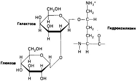
Collagen contains carbohydrate units covalently linked to hydroxylysine residues. Most commonly, this is a disaccharide of glucose and galactose (Fig. 9.4). The attachment of sugars occurs through the sequential action of galactosyltransferase and glucosyltransferase. These glycosylation Enzymes are specific for hydroxylysine residues in newly synthesized collagen that has not yet formed a triple helix. The number of carbohydrate units in tropocollagen depends on the tissue type. For example, in tendon collagen (type I) this number is 6, whereas in lens capsule collagen (type IV) it is 110.
Fig. 9.4. Carbohydrate components of collagen
9.5. The Structure of Tropocollagen Is a Triple-Stranded Helix
Let us now consider the conformation of the basic structural unit of type I collagen fiber.
As shown by electron microscopic and hydrodynamic studies, tropocollagen is a rod-shaped molecule 3000 A long and 15 A in diameter. It is one of the longest known Proteins. For comparison, tropocollagen is 60 times longer than the diameter of Chymotrypsin, and more than 2 times smaller in diameter. Each of the three polypeptide chains is helical (Fig. 9.5). In addition, these three helical chains wrap around each other to form a tight cable. Indeed, collagen fiber possesses remarkable strength: a force of 10 kg is required to break a fiber 1 mm in diameter.
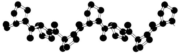
The helical nature of each chain in the three-stranded collagen fiber is well illustrated by the model compound poly-L-proline. This synthetic polypeptide has a helical conformation (Fig. 9.6), completely different from the α-Helix. It lacks Hydrogen Bonds. Instead, the helix is stabilized by steric repulsion between the pyrrolidine rings of the proline residues. As the polypeptide chain winds, the pyrrolidine rings are positioned as far apart as possible, forming a so-called type II trans-helix, which is much more extended than the tightly wound α-helix. The distance between two amino acid residues along the helix axis in poly-L-proline is 3.12 A, whereas in the α-helix it is 1.5 A. There are three amino acid residues per turn of the helix in poly-L-proline.
Fig. 9.5. Model of the collagen triple helix. Only α-carbon atoms are shown

Fig. 9.6. Model of the poly-L-proline helix (type II trans-helix)
A similar helix forms The basis of the three-dimensional Structure of each of the three tropocollagen chains
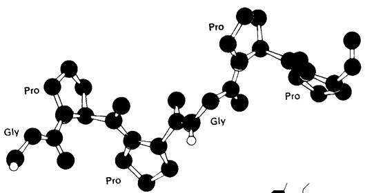
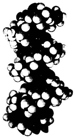
Let us now consider the conformation of an individual chain in the three-stranded Collagen helix (Fig. 9.7). Each chain is coiled similarly to poly-L-proline. These three helical chains then wrap around one another to form a superhelix (Fig. 9.8). The distance between two amino acid residues along the helix axis is 2.9 A, and there are nearly 3.3 residues per turn of the helix. The three chains are held together by hydrogen bonds. The peptide NH groups of glycine residues serve as hydrogen Donors, while the peptide CO groups of amino acid residues on other chains act as acceptors. The Hydrogen bonds are oriented nearly perpendicular to the long axis of the tropocollagen cable. The hydroxyl groups of hydroxyproline residues and Water molecules also participate in forming hydrogen bonds that stabilize the triple helix. A three-dimensional model of the collagen triple helix is shown in Fig. 9.9.
Fig. 9.7. Conformation of an individual chain in the collagen triple helix. Shown is a chain segment with the sequence -Gly-Pro-Pro-Gly-Pro-Pro
9.8. Skeletal model of the collagen triple helix. The repeating sequence -Gly-Pro-Pro is shown

Fig. 9.9. Three-dimensional model of the collagen triple helix
Last update: 06/08/2026
Editorial and Educational Adaptation: This material has been compiled based on the primary/original source text. The project team performed an editorial review, corrected technical inaccuracies, structured sections, and adapted the content for an educational format.
What was processed:
- elimination of formatting defects (OCR errors, structural breaks, corrupted characters);
- editorial organization of content;
- standardization of terminology in accordance with academic sources;
- verification of factual statements against the original source text.
All mentions of the author, publication year, and origin of the primary text have been preserved in accordance with the source.