BIOCHEMISTRY - L. Stryer - 1984
VOLUME 1
PART I. CONFORMATION AND DYNAMICS
CHAPTER 3. OXYGEN CARRIERS: MYOGLOBIN AND HEMOGLOBIN
3.6. A Rigid Heme Environment Ensures Reversible Oxygenation
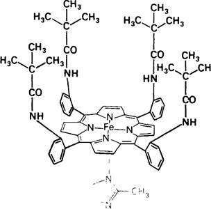
The oxygen-binding site accounts for only a small fraction of the total volume of the Myoglobin molecule. In fact, oxygen binds directly only to the iron atom within the heme. What is The Role of the polypeptide chain in oxygen binding and transport? To answer this question, let us examine how Oxygen binds to free heme. In aqueous solution, free ferroheme is capable of binding oxygen, but only momentarily. This is because ferroheme is rapidly oxidized in Water to ferriheme, which does not bind oxygen. The intermediate in this reaction is a sandwich compound formed by two Hemes with an O2 molecule trapped between them. In myoglobin, the heme is much less sensitive to oxidation because two myoglobin molecules are virtually incapable of associating to form a heme-O2-heme sandwich. The formation of such a complex is sterically hindered by the distal Histidine and other residues surrounding the sixth coordination position. The most compelling evidence that steric factors dictate The rate of heme oxidation comes from studies of specially synthesized model compounds. James Collman synthesized iron-porphyrin complexes surrounded by a picket fence of substituents (Fig. 3.19), which mimic the oxygen-binding sites in Myoglobin and Hemoglobin. In these compounds, one side of the porphyrin ring has a shielded pocket for O2 binding, while the other side remains open to bind a base. It turned out that when a substituted imidazole coordinates as the base, this complex (Fig. 3.20) acquires an oxygen affinity comparable to that of myoglobin. Furthermore, this picket-fence architecture stabilizes the ferrous form of the iron porphyrin, enabling it to undergo long-term reversible oxygen binding. The principal difference between this model compound and free heme lies in the presence of substituents that create the shielded pocket, thereby blocking the Formation of the dimeric heme-O2-heme sandwich.
Class="center">Fig. 3.18. Oxygen binding alters the visible absorption spectra of myoglobin and hemoglobin. Myoglobin and hemoglobin exhibit very similar visible Light absorption spectra.

Fig. 3.19. Schematic representation of a picket-fence iron porphyrin with bound O2. The steric hindrance of chemical substituents prevents two Porphyrins from approaching each other and forming the intermediate oxidation product.

Fig. 3.20. Structural formula of the picket-fence iron porphyrin.
Consequently, myoglobin provides a microenvironment for the prosthetic group that endows it with unique properties. It is a general rule that the function of a prosthetic group partly depends on its polypeptide surroundings. Indeed, the exact same heme performs a completely different function as part of cytochrome c, a component of the terminal Respiratory Chain in the Cell/35.html">Mitochondria of all aerobic organisms. In cytochrome c, heme reversibly transfers electrons rather than oxygen. Finally, heme plays an entirely different role in the enzyme catalase, where it decomposes hydrogen peroxide into water and oxygen.
3.7. The Presence of Distal Histidine Reduces Carbon Monoxide Binding
Carbon monoxide is toxic because, by binding to myoglobin or hemoglobin, it impairs Oxygen transport in the Organism. Heme has an exceptionally high affinity for CO. In aqueous solution, free heme binds CO roughly 25,000 times more strongly than O2. However, within hemoglobin or myoglobin, the affinity of heme for CO exceeds its affinity for O2 by only about 200-fold. How do these Proteins suppress heme's natural preference for carbon monoxide? The answer was provided by X-Ray Diffraction Analysis and Infrared Spectroscopy of CO and O2 complexes with myoglobin and model iron porphyrins. Iron porphyrins exhibit a very high affinity for CO; in their CO complexes, the Fe, C, and O atoms lie in a straight line. In the myoglobin-CO complex, however, the CO axis is tilted relative to the Fe—C bond. The linear alignment of CO with the Fe—C bond is primarily prevented by the presence of the distal histidine. On the other hand, the O2 axis is tilted relative to the Fe—O bond in both oxymyoglobin and model compounds. Consequently, under the Influence of the protein, CO coordinates to the iron at an angle to the Fe—C bond rather than colinearly. This tilted geometry, enforced by the globin, weakens the interaction between heme and CO while simultaneously creating optimal conditions for O2 binding.
The reduced affinity of myoglobin and hemoglobin for CO is of paramount importance for biological processes. Carbon monoxide posed a potential hazard long before the advent of industrialization, because CO is produced endogenously during heme degradation (Section 21.16). Thus, the generation of carbon monoxide within the body is inextricably linked to heme utilization. Endogenously produced CO blocks approximately 1% of the oxygen-binding sites in myoglobin and hemoglobin. Naturally, this level of inhibition is negligible. However, if the relative affinity of these proteins for CO were comparable to that of free iron porphyrins, endogenous carbon monoxide would cause severe poisoning. Nature resolved this issue through evolution by developing Hemoproteins that, due to steric factors, attenuate heme binding to CO while leaving O2 binding unaffected.
Last update: 06/08/2026
Editorial and Educational Adaptation: This material has been compiled based on the primary/original source text. The project team performed an editorial review, corrected technical inaccuracies, structured sections, and adapted the content for an educational format.
What was processed:
- elimination of formatting defects (OCR errors, structural breaks, corrupted characters);
- editorial organization of content;
- standardization of terminology in accordance with academic sources;
- verification of factual statements against the original source text.
All mentions of the author, publication year, and origin of the primary text have been preserved in accordance with the source.