BIOCHEMISTRY - L. Stryer - 1984
VOLUME 1
PART I. CONFORMATION AND DYNAMICS
CHAPTER 3. OXYGEN CARRIERS: MYOGLOBIN AND HEMOGLOBIN
3.5. The Oxygen-Binding Site in Myoglobin
The heme group is located in a crevice on the Myoglobin molecule. The highly polar propionate side chains of the heme are exposed on the molecular surface and exist in an ionized state at physiological pH. The remaining PARTS OF THE heme are embedded within the myoglobin interior, surrounded by nonpolar amino acid residues, with the exception of two histidines. The iron atom of the heme is covalently linked directly to one of these histidines, specifically residue F8 (Figs. 3.14 and 3.15). This residue, which occupies the fifth coordination position, is referred to as the proximal Histidine. The iron atom protrudes approximately 0.3 Å out of the porphyrin plane toward the histidine-F8 side. The oxygen-binding site is located on the opposite side of the plane at the sixth coordination position. A second histidine residue (E7), known as the distal histidine, is situated nearby. The distal histidine residue E7 is not bonded to the heme. Figure 3.16 shows a section of the electron density map encompassing the heme region.
Class="center">Fig. 3.14. Schematic representation of the oxygen-binding site in myoglobin. The fifth coordination position is occupied by histidine F8 (proximal histidine); oxygen binds at the sixth coordination position; histidine E7 (distal histidine) is located near the sixth coordination position
Fig. 3.15. Model of the oxygen-binding site in myoglobin, showing the heme, proximal histidine (F8), and distal histidine (E7)
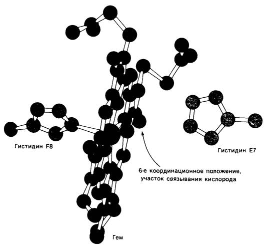
Fig. 3.16. Section of the myoglobin electron density map near the oxygen-binding site. The electron-dense region extending along the lower part of the map is the E helix

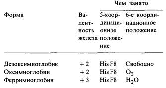
Three physiologically relevant forms of myoglobin have been studied: deoxymyoglobin, oxymyoglobin, and metmyoglobin. They are conformationally very similar in every respect except for the sixth coordination position (Table 3.1). In metmyoglobin, this position is occupied by a Water molecule; in deoxymyoglobin, it is vacant; in oxymyoglobin, it is occupied by oxygen. The binding to O2 is slanted relative to the iron-oxygen bond axis (Fig. 3.17). Upon oxygenation, the iron atom moves inward toward the heme plane by approximately 0.2 Å. It is important to note that the heme is therefore not a rigid Structure. Furthermore, the Displacement of the iron atom upon oxygenation plays a pivotal role in Hemoglobin function, as will be discussed in the next chapter (Sec. 4.11).
Fig. 3.17. Tilt and orientation of the oxygen molecule bound to the iron atom in oxymyoglobin. The angle between the O2 axis and the Fe — O bond is 121°
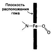
Table 3.1. The heme environment
The heme makes contact with amino acid residues originating from different segments of the linear sequence, notably residues C4 and H14, which correspond to Amino Acids 39 and 138 in the primary sequence. This demonstrates that the heme-binding pocket is largely a product of three-dimensional folding.
Last update: 06/08/2026
Editorial and Educational Adaptation: This material has been compiled based on the primary/original source text. The project team performed an editorial review, corrected technical inaccuracies, structured sections, and adapted the content for an educational format.
What was processed:
- elimination of formatting defects (OCR errors, structural breaks, corrupted characters);
- editorial organization of content;
- standardization of terminology in accordance with academic sources;
- verification of factual statements against the original source text.
All mentions of the author, publication year, and origin of the primary text have been preserved in accordance with the source.