BIOCHEMISTRY - L. Stryer - 1984
VOLUME 1
PART I. CONFORMATION AND DYNAMICS
CHAPTER 3. OXYGEN CARRIERS—MYOGLOBIN AND HEMOGLOBIN
3.4. The Structure of Myoglobin is Characterized by Compactness and a High Degree of α-Helicity
The high-resolution electron density map of Myoglobin, obtained two years later, yielded a tremendous amount of detail regarding the Cell/13.html">Protein Structure. Out of the 1,260 atoms—that is, all atoms except hydrogen—the positions of 120 atoms were determined with an accuracy exceeding 0.3 Å. The overall arrangement of the main chain and the heme group is shown in Fig. 3.12. The structure of myoglobin exhibits the following features:
1. The myoglobin molecule is extremely compact. Its dimensions are 45 x 35 x 25 Å. There is virtually no free space inside the molecule.
Class="center">Fig. 3.11. Low-resolution model of myoglobin
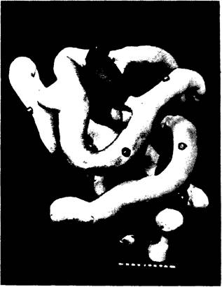
Fig. 3.12. High-resolution model of myoglobin. Only α-carbon atoms are shown. The heme is colored red
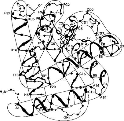
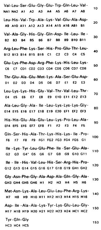
2. About 75% of the main chain is in the α-helical conformation. All α-helices are right-handed. There are 8 main helical regions, designated by the first 8 letters of the Latin alphabet: A, B, C, ..., H. The first amino acid residue in helix A is designated A1, the second A2, and so on (Fig. 3.13). Between the helical regions, There are five non-helical segments (designated, for example, CD if the segment is located between helices C and D). There are two additional non-helical segments: two amino acid residues at the N-terminus of the molecule (designated NA1 and NA2) and five residues at the C-terminus (designated HC1 to HC5, respectively).
Fig. 3.13. Amino Acid Sequence of sperm whale myoglobin. Numbers and letters beneath each residue indicate its position within the α-helices or in the non-helical segments between them. For example, B4 denotes the fourth residue in helix B; EF7 denotes the 7th residue in the non-helical segment between helices E and F
3. Not all the factors that determine the termination of a helical region have been fully identified. However, it is known that Proline residues play an important role in this process. Proline cannot be incorporated into an α-Helix (except possibly at the very end), because its five-membered pyrrolidine ring simply cannot fit into it. Myoglobin contains four proline residues and eight α-helix terminations. Clearly, other factors are also involved in determining helix termination. For instance, in certain cases, the α-helix is interrupted when the OH group of Serine or Threonine interacts with the carbonyl group of the main chain.
4. The peptide groups have a planar structure. The carbonyl group is in a *trans* conformation relative to the backbone NH group. Bond Lengths and angles correspond to those found in dipeptides and related compounds.
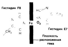
5. The interior and exterior of the molecule are clearly distinguishable. The interior consists almost entirely of nonpolar residues such as leucine, valine, Methionine, and phenylalanine, and lacks side chains of glutamic acid, aspartic acid, glutamine, asparagine, Lysine, or Arginine. Amino acid residues possessing both polar and nonpolar parts—specifically threonine, Tyrosine, and Tryptophan—are oriented with their nonpolar portions facing inward. There are only two polar residues inside the myoglobin molecule: two Histidine residues located in the Active Site that play a key role in its functional activity. Both polar and nonpolar amino acid residues are located on the outer surface of the molecule.
Last update: 06/08/2026
Editorial and Educational Adaptation: This material has been compiled based on the primary/original source text. The project team performed an editorial review, corrected technical inaccuracies, structured sections, and adapted the content for an educational format.
What was processed:
- elimination of formatting defects (OCR errors, structural breaks, corrupted characters);
- editorial organization of content;
- standardization of terminology in accordance with academic sources;
- verification of factual statements against the original source text.
All mentions of the author, publication year, and origin of the primary text have been preserved in accordance with the source.