Protein Chemistry. Structure, Properties, and Research Methods - Shendryk A.N. 2022
Protein Structure
Behavior of Proteins in Solutions
Acid-Base Properties of Proteins
The acid-base properties of native Proteins are determined primarily by the presence and number of ionizable R-groups in their amino acid residues. The two terminal Functional groups of the peptide chain—the amino and carboxyl groups—have practically no effect on the acid-base Properties of the protein molecule as a whole.
The degree of ionization of the functional groups in the R-residues of Amino Acids comprising a protein molecule is determined not only by its Primary Structure (Amino Acid Sequence), but also by higher-order structures. Therefore, it is impossible to accurately predict the acid-base properties and net charge of a protein molecule based solely on its known amino acid sequence.
Let us consider the potentiometric titration curve of Ribonuclease.
Class="center">
Fig. 2.6 Potentiometric titration curve of ribonuclease
The peptide chain of this protein molecule is folded into a dense globule consisting of 124 amino acids with a known sequence. In addition to the terminal residues with -NH2 and -COOH groups, the peptide chain contains 34 ionizable R-groups, most of which exhibit basic properties. Consequently, The titration curve has a relatively "smoothed" shape compared to titration curves of simple compounds, such as a dibasic acid (see fig.).
As can be seen from the titration curve of ribonuclease (see Fig. 2.6), the isoelectric point for this protein is shifted toward higher pH values, meaning that the molecule as a whole is characterized by basic rather than acidic properties. Very similar results were obtained upon titration of ß-lactoglobulin, egg albumin, and A number of other Globular proteins. Based on these data, it was concluded that the vast majority of ionizable R-groups incorporated into the peptide chain are accessible to acid-base titration. This Conclusion is in good agreement with X-Ray Diffraction data showing that almost all ionogenic R-groups in globular proteins are located on the outer surface of the molecule, while most nonpolar (hydrophobic) groups are concealed within the core. Nevertheless, in some globular proteins, one or more ionizable R-groups fail to titrate. It is assumed that they are either located inside the globule or involved in The formation of stable Hydrogen Bonds. For instance, in Myoglobin, 5 out of its 11 Histidine R-groups can be titrated only after Protein Denaturation.
Proteins, much like amino acids, are characterized by the presence of isoelectric points—pH values at which their molecules carry a net zero charge. The method of isoelectric focusing of proteins is based on this property. The positions of isoelectric points are determined by the number and type of amino acids with ionizable R-groups that make up the protein. The higher the content of basic R-groups (e.g., Lys, Arg) in the protein peptide chain, the higher the isoelectric point relative to pH = 7. Conversely, the more acidic R-residues (Asp, Glu, etc.) the protein molecule contains, the lower the isoelectric point. For the majority of globular proteins, isoelectric points lie within the pH range of 4.5 - 6.5 (see Table 2.1).
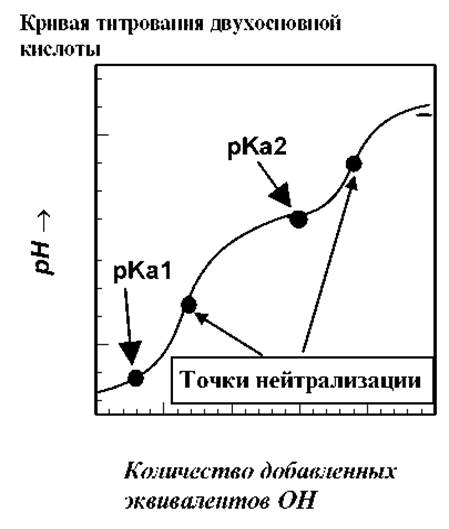
Table 2.1 Positions of isoelectric points for certain proteins
|
Protein |
Isoelectric point pH |
< 1.0 |
|
|
Egg albumin |
4.6 |
|
Serum albumin |
4.9 |
|
Urease |
5.0 |
|
ß-Lactoglobulin |
5.2 |
|
y1-Globulin |
5.6 |
|
6.8 |
|
|
Myoglobin |
7.0 |
|
Chymotrypsinogen |
9.5 |
|
Cytochrome c |
10.65 |
|
11.0 |
Potentiometric titration curves make it possible to determine the magnitude and sign of the net charge of a protein molecule at a given pH value. At a pH above the isoelectric point, the protein molecule is negatively charged and will migrate toward the anode in an electric field. As the pH increases, the magnitude of the negative charge of the protein molecule will increase in accordance with the shape of the titration curve. At a pH below the isoelectric point, the protein molecule is positively charged and migrates toward the cathode.
The profile of the titration curve strongly depends on the presence of neutral salts in the solution, as they affect the ionization degree of individual R-groups in various ways. Furthermore, proteins tend to bind inorganic cations (such as Cu2+, Mg2+) and anions (e.g., chloride and phosphate ions). For this reason, THE POSITION OF the isoelectric point for the same protein can vary significantly depending on the COMPOSITION OF THE medium.
Last update: 06/08/2026
Editorial and Educational Adaptation: This material has been compiled based on the primary/original source text. The project team performed an editorial review, corrected technical inaccuracies, structured sections, and adapted the content for an educational format.
What was processed:
- elimination of formatting defects (OCR errors, structural breaks, corrupted characters);
- editorial organization of content;
- standardization of terminology in accordance with academic sources;
- verification of factual statements against the original source text.
All mentions of the author, publication year, and origin of the primary text have been preserved in accordance with the source.