Biochemistry: The Chemical Reactions of Living Cells, Volume 3 - D. Metzler 1980
Biochemical Genetics and the Synthesis of Nucleic Acids and Proteins
Translation of Genetic Information. Protein Synthesis
Chemical Composition of Ribosomes
The protein-synthesizing system of E. coli includes 15,000 Ribosomes, which account for one-fourth of the total Cell mass. Elucidating the Structure and operating mechanism of these tiny molecular machines, which appear merely as indistinct dots under an Electron microscope, is one of the main research directions in modern molecular biology [88].
The mass of an E. coli ribosome is approximately 2.7∙106 daltons; about 65% of its weight is RNA, and the remaining 35% is protein. In Eukaryotic Cells, the ribosomal mass is about 1.6 times greater than in bacterial cells (4.3∙106 daltons). Under certain conditions, specifically at low concentrations of Mg2+ ions, intact ribosomes (70S ribosomes in Bacteria) dissociate into two unequal subunits — the 30S and 50S ribosomal subunits. The 50S subunit is roughly twice as large as the 30S subunit and contains two RNA molecules (23S and 5S) (Table 15-4). The smaller (30S) subunit contains a single 16S rRNA molecule, whose polynucleotide chain comprises 1,700 NUCLEOTIDES; its length, if fully stretched out, can exceed 500 nm. The nucleotide sequence of this RNA has been completely deciphered.
Class="center">Table 15-4 Mass of E. coli ribosomes
|
Particle |
Ribosome, daltons (x106) |
RNA, daltons (x105) |
Protein (by difference) daltons (x103) |
|
50S subunit 30S subunit 70S subunit |
∼1.8 ∼0.9 ∼2.7 |
∼1.1 (23S) ∼0.04 (5S) ∼0.56 (16S) ∼1.7 |
∼0.7 ∼0.3 ∼1.0 |
In addition to densely packaged RNA molecules, the 30S subunit contains approximately 21 protein molecules that vary in Amino Acid Composition and sequence (Table 15-5). Many of these Proteins (designated as S1, S2, S3, etc.) have a relatively low molecular weight. Furthermore, many exhibit strongly basic properties, containing A large number of Lysine and Arginine residues that undoubtedly mediate the interaction of these proteins with RNA molecules. At the same time, the 30S subunit also contains several acidic and neutral proteins. The 50S ribosomal subunit contains ~34 different proteins, and a single subunit may harbor multiple copies of the same protein type. The protein composition of ribosomes can undergo variations, making its precise determination quite difficult. Most proteins (usually referred to as structural units) are present in a 1:1 ratio. Other proteins may be absent in certain ribosomes. Similarly, additional copies of some subunits may be contained in only a fraction of the ribosomes. During Protein Synthesis, A number of other proteins can temporarily associate with functioning ribosomes.
Determining the exact size and shape of ribosomes is a challenging task. Currently, the diameter of bacterial ribosomes is estimated to be approximately 22 nm, with a particle length possibly reaching 30 nm. Eukaryotic ribosomes are roughly 1.17 times larger in linear dimensions and contain a significantly greater number of proteins—about thirty in the small subunit and about forty in the large subunit [89]. However, there is reason to believe that the number of functionally essential proteins in eukaryotic ribosomes is the same as in E. coli ribosomes [90]. Interestingly, the proteins of eukaryotic ribosomes (as well as rRNA molecules) are considerably larger than those of bacterial ribosomes. Mitochondrial ribosomes resemble bacterial ones in some respects, but are larger and contain approximately 66% protein (whereas E. coli ribosomes contain only 35% protein).
Abundant evidence indicates that ribosomal proteins exist as compact molecules whose surfaces are maximally accessible to added Reagents. RNA molecules are also largely accessible to external influences. About 50% of the total ribosomal mass is in a hydrated state; thus, ribosomes are structures that can be relatively easily penetrated by solvent. Most of the RNA (perhaps 60–70%) is folded into base-paired loops, much like tRNA. Numerous experiments have been conducted to elucidate the physical principles underlying the binding of different ribosomal subunits to each other. These studies revealed that proteins S4 and S20 bind directly to 16S rRNA near its 5'-end, proteins S8 and S15 near the center, and protein S7 near the 3'-end. It has been suggested that protein S4 plays a particularly crucial role in ribosome Organization [88]. Methods have now been developed to fully dissociate the 30S and 50S ribosomal subunits of E. coli [91, 92] into individual proteins and RNA, followed by their reconstitution into functionally active ribosomes. Such experiments have established that the order of protein addition is critical for proper reconstruction. These findings suggest that in addition to proteins S4, S7, S15, and S20, proteins S9, S13, and S17 also bind to 16S rRNA, while protein S16 binds to proteins S4 and S20 [93]. The remaining proteins are not required for structural restoration of the ribosome, but are essential for its function; these include proteins S3, S10, S12, S14, and S19. No enzymatic activity has been detected in most ribosomal proteins, though it is quite possible they possess other, as yet undiscovered catalytic properties.
Table 15-5 Ribosomal proteins
|
Proteins of 30S ribosomal subunits |
Proteins of 50S ribosomal subunits |
||||
|
Designation |
Mol. weight |
Bindinga |
Designation |
Mol. weight |
Bindinga |
|
S1 |
65 000 |
L1 |
22000 |
||
|
S2 |
27 000 |
L2 |
28 000 |
+ |
|
|
S3 |
28 000 |
L3 |
23 000 |
||
|
S4 |
25 000 |
+ |
L4 |
28 500 |
|
|
S5 |
21 000 |
L5 |
17 500 |
||
|
S6 |
17 000 |
L6 |
21 000 |
+ |
|
|
S7 |
26 000 |
+ |
L7 |
15 500 |
|
|
S8 |
16 000 |
+ |
L8 |
19 000 |
|
|
S9 |
17 500 |
L9 |
|||
|
S10 |
17 000 |
L10 |
21 000 |
||
|
S11 |
L11 |
19 000 |
|||
|
S12 |
17 000 |
L12 |
15 500 |
||
|
S13 |
14 000 |
L13 |
20 000 |
||
|
S14 |
15 000 |
L14 |
18 500 |
||
|
S15 |
13 000 |
+ |
L15 |
17 000 |
|
|
S16 |
13 000 |
L16 |
22 000 |
+ |
|
|
S17 |
10 000 |
L17 |
15 000 |
+ |
|
|
S18 |
12 000 |
L18 |
17 000 |
+ |
|
|
S19 |
14 000 |
L19 |
17500 |
+ |
|
|
S20 |
13 000 |
+ |
L20 |
16 000 |
+ |
|
S21 |
13 000 |
L21 |
14 000 |
||
|
Total |
405 000 |
L22 |
17 000 |
||
|
L23 |
12 500 |
+ |
|||
|
L24 |
14500 |
+ |
|||
|
L25 |
12 500 |
+ |
|||
|
L26 |
12 500 |
||||
|
L27 |
12 000 |
||||
|
L28 |
15 000 |
||||
|
L29 |
12 000 |
||||
|
L30 |
10 000 |
||||
|
L31 |
|||||
|
L32 |
|||||
|
L33 |
9 000 |
||||
|
L34 |
|||||
|
Total 549 000 |
' |
||||
The plus sign (+) denotes direct binding to ribosomal RNA.
It has been shown that proteins L5, L18, and L25 of the 50S subunit specifically bind to the 5S rRNA molecule, whose base sequence has been established (Fig. 15-12). It is also known that the L5-L18-L25-5S rRNA complex binds to the oligonucleotide TpψpCpGp. Consequently, it can be hypothesized that 5S rRNA interacts with the TψC-arm of the tRNA molecule when the latter binds to the ribosome. Furthermore, it was found that L18+L5 (or L25) mediate the binding of 5S rRNA to 23S rRNA. Table 15-4 lists the remaining Proteins of the 50S ribosomal subunits that bind to RNA.
Numerous experiments are currently being conducted using cross-linking reagents between protein molecules. In particular, bifunctional compounds capable of covalently binding to two different —SH or —NH2 groups are employed [93–95]. This approach has made it possible to identify the following cross-linked pairs [93, 95]: S2-S3, S4-S6, S4-S8, S4-S9, S4-S12, S5-S8, S5-S9, S7-S8, S7-S9, S11-S18, S13-S19, and S18-S21. Another approach consists in producing specific Antibodies against individual ribosomal proteins and studying their binding sites On the surface of ribosomal subunits using an electron microscope [96, 97]. This method has established the localization of many proteins on The surface of both 30S and 50S subunits (Fig. 15-13). In several cases, antibodies against a specific protein bound to multiple sites at once. The fact that the binding sites for antibodies against proteins S2, S12, S15, and S18 are separated by 8–19 nm indicates that these proteins exist in an extended, fibrillar conformation in the 30S subunit (Fig. 2-12). Based on similar data, protein S4 is also believed to have an extended shape, reaching a length of 17 nm [95], which enables this protein to form a large number of cross-linked pairs. All of this demonstrates that the ribosome is an extraordinarily complex machine.
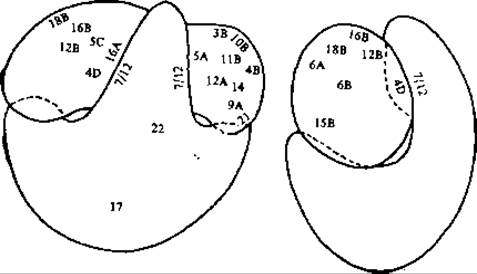
FIG. 15-13. Two projections of the E. coli 70S ribosome. Numbers indicate the binding sites of specific antibodies against particular ribosomal proteins (numbering is the same as in Table 15-5). The letters S and L are omitted, as the diagram clearly shows which subunit houses the protein. In cases where more than one binding site is found for a single antibody, these sites are designated by the letters A, B, C, and D. Many other identified binding sites would be visible in other projections [97].
Last update: 06/08/2026
Editorial and Educational Adaptation: This material has been compiled based on the primary/original source text. The project team performed an editorial review, corrected technical inaccuracies, structured sections, and adapted the content for an educational format.
What was processed:
- elimination of formatting defects (OCR errors, structural breaks, corrupted characters);
- editorial organization of content;
- standardization of terminology in accordance with academic sources;
- verification of factual statements against the original source text.
All mentions of the author, publication year, and origin of the primary text have been preserved in accordance with the source.