Human Biochemistry Volume 2 - Murray R. 1993
Biochemistry of Intracellular and Intercellular Communications
Sex Gland Hormones
Biosynthesis and Metabolism of Testicular Hormones
Synthesis
Testicular androgens are synthesized by Leydig Cells within the interstitial tissue. These cells contain virtually all of the testicular 3β-hydroxysteroid dehydrogenase, the enzyme that catalyzes the key step in testosterone Biosynthesis.
A. Biosynthetic Pathways
1. Testosterone serves as the immediate precursor for sex Steroids, much as Cholesterol serves as the precursor for adrenal corticosteroids. As in the Adrenal Glands, the rate-limiting step is the Cleavage of the cholesterol side chain. The conversion of cholesterol to pregnenolone occurs identically in the adrenal glands, Ovaries, and Testes; however, in the latter two Tissues, this reaction is stimulated by LH rather than ACTH.
The conversion of pregnenolone to testosterone involves five Enzymes: 1) 3β-hydroxysteroid dehydrogenase (3β-HSD); 2) ∆5,4-isomerase; 3) 17α-hydroxylase; 4) C17-20-lyase; and 5) 17β-hydroxysteroid dehydrogenase (17β-HSD). The corresponding sequence of reactions, known as the progesterone (or ∆4) pathway, is shown on the right in Fig. 50.1. Alternatively, the conversion of pregnenolone to testosterone can proceed via the dehydroepiandrosterone (or ∆5) pathway (Fig. 50.1, left). In human testes, the ∆4 pathway appears to predominate. It is important to bear in mind, however, that human testicular tissue is largely inaccessible for research, and most studies elucidating these pathways have been conducted in animals; significant species variations may therefore exist.
The five aforementioned enzymes are localized in the microsomal fraction of rat testes, with a close functional coupling observed between the activities of 3β-HSD and ∆5,4-isomerase, as well as between 17α-hydroxylase and C17-20-lyase. These enzyme pairs are depicted in the overall reaction sequence in Fig. 50.1 and in the scheme of androgen biosynthetic pathways within testicular microsomal membranes in Fig. 50.2. The latter figure illustrates the influx of various substrates for testosterone biosynthesis into the microsomes and their sequential incorporation into the ∆4 pathway chain. Because there are four potential substrates for what appears to be a single 3β-HSD, numerous alternative pathways exist. The pathway selected in any given instance likely depends on local substrate concentrations near the respective enzymes, which may be modulated by substrate compartmentalization within the microsomal membrane.
2. Other Testicular Hormones. Dihydrotestosterone (DHT) is formed from testosterone via the reduction of ring A by the enzyme 5α-reductase. Daily secretion of DHT by human testes is approximately 50–100 µg; however, the vast majority of circulating DHT results from peripheral conversion (see below).
The testes also produce small yet significant amounts of 17β-estradiol (E2), a female sex hormone. Most of the E2 produced in males stems from the peripheral aromatization of testosterone and androstenedione. E2 synthesis is believed to involve Leydig cells, Sertoli cells, and the seminiferous tubules. The Physiological Role of E2 in males remains unclear, though it may participate in The regulation of FSH. Abnormally high plasma E2 levels and an altered free E2-to-testosterone ratio are characteristic of pubertal or postpubertal gynecomastia, chronic Liver disease, and hyperthyroidism.
Class="center">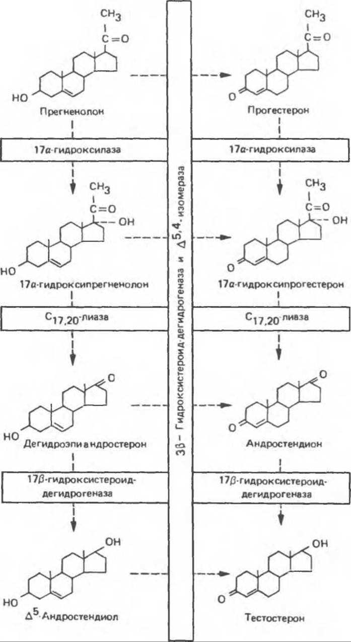
Fig. 50.1. Pathways of testosterone biosynthesis. The dehydroepiandrosterone (or ∆5) pathway is shown on the left, and the progesterone (or ∆4) pathway on the right.
B. Age-Related Changes in Testicular Hormone Production
In rat fetuses and newborns, testosterone predominates; however, shortly after birth, the testes begin producing exclusively androsterone. The capacity to synthesize testosterone is restored at Puberty and persists throughout life. Comparable findings have been obtained in other species, and it is highly likely that similar age-related shifts occur in humans.

Fig. 50.2. Schematic representation of androgen biosynthesis in the testicular microsomal membrane. The membrane is depicted as a horizontal line, which may reflect its appearance in situ; however, in microsomal preparations, it forms vesicles. A = androstenedione, T = testosterone. (Reproduced, with permission, from DeGroot L. J. Endocrinology, Vol. 3, Grune and Stratton, 1979.)
Secretion and Transport
Although several steroids are present in testicular venous Blood, the principal steroid secreted by the adult Testis is testosterone. Normal daily testosterone secretion in men is approximately 5 mg. The secretion of testicular steroids does not appear to be regulated; like other Steroid Hormones, testosterone is apparently secreted as it is synthesized.
Table 50.1. Hormone Binding by Sex Hormone-Binding Globulin (SHBG)
|
Bound Steroids |
Unbound Steroids |
|
Testosterone |
Conjugated androgens |
|
17β-Estradiol |
17α-Testosterone |
|
Dihydrotestosterone |
Dehydroisoandrosterone |
|
Other 17β-hydroxy steroids |
Cortisol |
|
Estrone |
Progesterone |
The plasma of most mammals, including humans, contains a β-globulin that specifically binds testosterone with relatively high affinity and limited capacity (Table 50.1). This protein, commonly referred to as sex hormone-binding globulin (SHBG) or testosterone-estradiol-binding globulin (TEBG), is synthesized in the liver. Its production is enhanced by estrogens (serum SHBG concentrations in women are twice those in men), in certain liver diseases, and in hyperthyroidism, whereas it is decreased by androgens, Aging, and hypothyroidism. Many of these factors also influence the synthesis of corticosteroid-binding globulin (see Chapter 48) and thyroid-binding globulin (see Chapter 46). Because SHBG and albumin bind 97% to 99% of circulating testosterone, only a minute fraction exists in the blood in a free (biologically active) form. The primary function of SHBG is likely to restrict the concentration of free testosterone in serum. Because testosterone binds to SHBG with a higher affinity than estradiol does (Table 50.2), alterations in SHBG concentration affect free testosterone levels to a greater extent than free estradiol levels. An elevation in SHBG concentration drives up the free estradiol-to-testosterone ratio. This phenomenon occurs during aging, liver cirrhosis, and hyperthyroidism, thereby contributing to the signs and symptoms of "feminization" characteristic of these conditions.
Table 50.2. Approximate Steroid Affinities for Plasma Binding Proteins
|
Hormone |
SHBG 1) |
CBG 1) |
|
Estradiol |
5 |
> 10 |
|
Estrone |
> 10 |
> 100 |
|
Androstenedione |
... |
... |
|
Testosterone |
2 |
> 100 |
|
Dihydrotestosterone |
1 |
> 100 |
|
Progesterone |
> 100 |
2 |
|
Cortisol |
> 100 |
3 |
1) Affinity is expressed as moles × 109. (Adapted from Siiteri R. K., Febres F. Ovarian hormone synthesis, Circulation and Mechanisms of action. Page 1401. In: Endocrinology, Vol. 3. DeGroot L. J. (editor). Grune and Stratton, 1979.)
Peripheral METABOLISM and Excretion
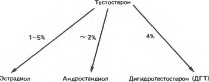
A. Metabolic Pathways. The metabolic transformation of testosterone proceeds via two main pathways. One involves oxidation at the 17-position, and the other involves reduction of the ring A double bond and the 3-keto group1. The first pathway, active in numerous tissues including the liver, yields 17-ketosteroids that are generally devoid of activity or possess significantly weaker activity than the parent compound. The second pathway, which is less efficient, occurs predominantly in target tissues and leads to The formation of the active metabolite DHT, as well as estradiol and androstanediol. Etiocholanolone and androsterone are 5β-reduced androgen metabolites.
B. Testosterone Metabolites. The most important metabolite of testosterone is DHT, which represents the active form of the hormone and is found in many tissues, including the Seminal Vesicles, Prostate Gland, external genitalia, and certain Regions of the Skin. In the plasma of adult men, DHT levels are roughly one-tenth those of testosterone, with approximately 400 µg produced daily (compared to 5 mg of testosterone). The conversion of testosterone to DHT is catalyzed by a NADPH-dependent 5α-reductase.
Thus, testosterone can be considered a prohormone for two reasons: first, it is converted into a more active compound, dihydrotestosterone, and second, this conversion occurs primarily in tissues located outside the testes. A small fraction of testosterone is aromatized to form estradiol, which is particularly important for the Brain, where these hormones are involved in shaping the Sexual Behavior of animals. Androstanediol, another highly active androgen, is also formed from testosterone.
1 According to the new nomenclature, it should be written as oxogroup, oxosteroids, etc., but since the term "ketosteroids" is standard in medical literature, we will stick to this name here.—Trans. note.
The main 17-Ketosteroid metabolites of testosterone—androsterone and etiocholanolone—are conjugated in the liver with glucuronide and sulfate to form Water-soluble excretory compounds. The Quantitative determination of urinary 17-ketosteroids was previously used as a test for androgenic activity. Now, however, it has been established that this indicator poorly reflects the in vivo hormonal status.
Last update: 06/08/2026
Editorial and Educational Adaptation: This material has been compiled based on the primary/original source text. The project team performed an editorial review, corrected technical inaccuracies, structured sections, and adapted the content for an educational format.
What was processed:
- elimination of formatting defects (OCR errors, structural breaks, corrupted characters);
- editorial organization of content;
- standardization of terminology in accordance with academic sources;
- verification of factual statements against the original source text.
All mentions of the author, publication year, and origin of the primary text have been preserved in accordance with the source.