Principles of Biochemistry Volume 3 - A. Lehninger 1985
Molecular Mechanisms of Genetic Information Transfer
DNA Replication and Transcription
Replication requires the physical separation of the strands of the parental double-stranded DNA
We have already seen that circular double-stranded DNA replicates simultaneously in both directions, causing two Replication forks to move toward each other along the circular chromosome. Recalling that the DNA double helix is a tightly wound Structure with the coding bases buried inside, it becomes clear that for DNA-replicating Enzymes to “read” the template nucleotide sequence, the parental DNA strands must be separated, at least locally. Even if Bacterial DNA is negatively supercoiled—meaning it is already slightly unwound—it must undergo further unwinding as the Replication fork advances.
The unwinding of The Double Helix and the stabilization of the separated strands to allow for replication are carried out by several specialized Proteins (Fig. 28-13). Enzymes known as helicases unwind short stretches of DNA directly ahead of the replication fork. This unwinding process requires energy: the Separation of each base pair consumes the energy derived from the Hydrolysis of two ATP molecules to ADP and inorganic phosphate. As soon as a short segment of DNA is unwound, several molecules of single-stranded DNA-binding protein (SSB) bind tightly to each separated strand, preventing complementary base pairing and reannealing. This makes The nucleotide sequences of the DNA strands accessible to the replication machinery. DNA polymerase can directly extend the leading strand by adding new NUCLEOTIDES to its 3'-end. Other specific proteins assist primase in gaining access to the lagging-strand template, enabling primase to bind and synthesize RNA primers for Okazaki fragments. DNA unwinding is one of the most intriguing yet challenging problems in METABOLISM/36.html">DNA replication; much remains to be understood about how the living Cell solves this mechanical and biochemical puzzle. The key proteins required for DNA replication are shown in Fig. 28-13.
Class="center">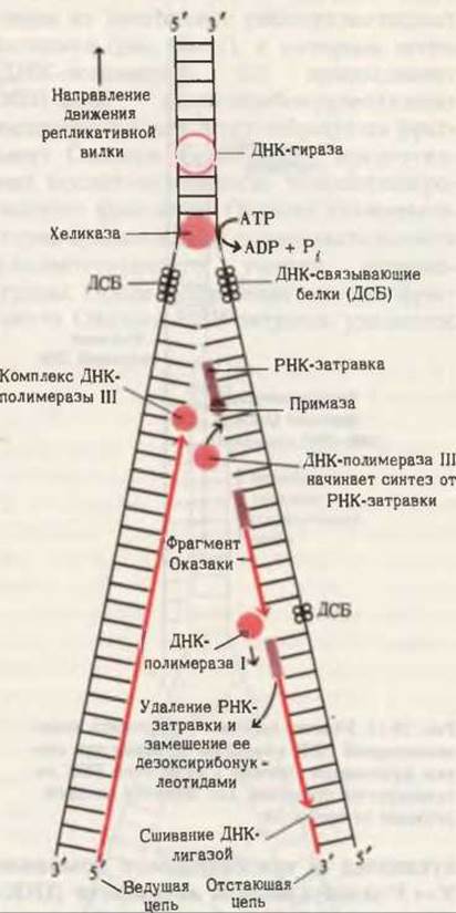
Fig. 28-13. Schematic representation of the MAIN STAGES OF DNA replication. The precise Site of Action of DNA gyrase remains somewhat uncertain.
The rapid unwinding of parental DNA strands during replication (at 4,500 rpm) poses another problem: in the absence of a specialized swivel mechanism, the entire chromosome ahead of the replication fork would have to rotate at the same high speed. It is thought that The Cell avoids this by utilizing a swivel point in the DNA (presumably located just ahead of the replication fork), which confines the high-speed rotation to only a short segment of DNA. This is achieved through a transient nick in one of the DNA strands, which is rapidly and precisely resealed after one or more rotations. These transient breaks and reunions are catalyzed by enzymes known as topoisomerases. In prokaryotes, this topoisomerase is called DNA gyrase. Not only does this enzyme allow the DNA to rotate, but it also actively twists the DNA in a direction that favors the unwinding of the template strands near the replication fork. Thus, gyrase assists helicase in unwinding DNA for replication. The gyrase-mediated supercoiling of DNA, coupled with the hydrolysis of ATP to ADP and Pi, maintains the supercoiled state of the chromosome. Thanks to gyrase, all circular DNA molecules in bacterial Cells are kept in a supercoiled form (Fig. 28-14).
As the replication machinery seals the nicks in the lagging strand, it progresses along the replicating DNA. The two new strands automatically pair with their complementary template strands, forming two daughter Double helices, each containing one parental and one newly synthesized strand. The formation of these new helices requires neither energy input nor the action of any “twisting” enzyme.

Fig. 28-14. Formation of negative supercoils by DNA gyrase. The energy required for this process is released through the hydrolysis of ATP. DNA gyrase introduces a break in one of the DNA strands, passes the intact strand through the resulting gap, and then reseals the broken ends.
Last update: 06/08/2026
Editorial and Educational Adaptation: This material has been compiled based on the primary/original source text. The project team performed an editorial review, corrected technical inaccuracies, structured sections, and adapted the content for an educational format.
What was processed:
- elimination of formatting defects (OCR errors, structural breaks, corrupted characters);
- editorial organization of content;
- standardization of terminology in accordance with academic sources;
- verification of factual statements against the original source text.
All mentions of the author, publication year, and origin of the primary text have been preserved in accordance with the source.