Biochemistry and Molecular Biology - Belyasova N.A. 2002
Molecular Foundations and Mechanisms of Heredity
Gene Expression
Translation of the Genetic Code
Translation is The process of mRNA decoding, whereby the information encoded in The nucleotide sequence of an mRNA molecule is translated into the Amino Acid Sequence of a polypeptide. The decoding of mRNA proceeds in the 5' → 3' direction. The process of translation involves the following stages:
2) tRNA aminoacylation;
3) translation proper.
Amino acid activation. This is the process wherein an amino acid is attached via its carboxyl group to the a-phosphate of ATP with the participation of a specific aminoacyl-tRNA synthetase (Fig. 3.10). The reaction is accompanied by the release of inorganic pyrophosphate and The formation of aminoacyl-adenylate (AA-AMP). Aminoacyl-adenylate is highly reactive and is stabilized by tight binding to the enzyme. This process is characterized by high Specificity: each amino acid has its own dedicated enzyme(s).
Class="center">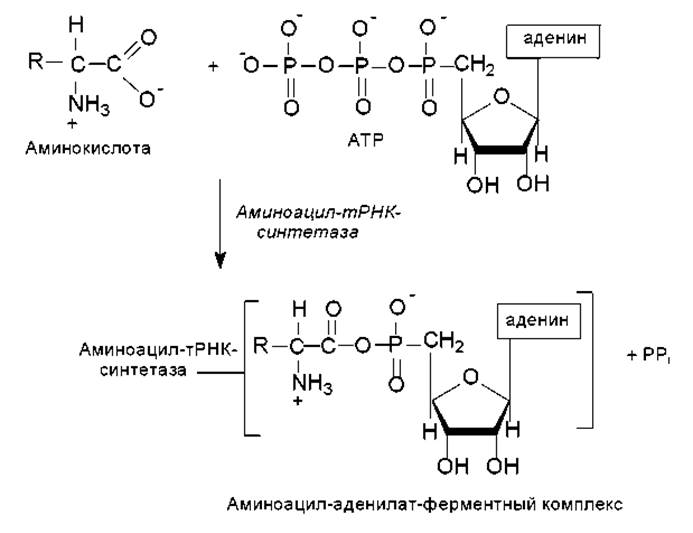
Fig. 3.10. Amino acid activation during translation
tRNA aminoacylation. This involves The transfer of the aminoacyl group from the enzyme-bound aminoacyl-adenylate to the 2'- or 3'-OH group of the terminal ribose of tRNA within the acceptor stem (Fig. 3.11).
A key feature of the reaction leading to tRNA aminoacylation is the Specificity of the Enzymes involved. The attachment of each of the 20 Amino Acids found in Proteins to its corresponding tRNA is catalyzed by a specific aminoacyl-tRNA synthetase. The enzyme must distinguish one amino acid from the other 19 and transfer it to one or more isoacceptor tRNAs out of approximately 75 other available tRNAs. It should be emphasized that many amino acids share a high degree of structural similarity (e.g., leucine, valine, and isoleucine; valine and Threonine; aspartic and glutamic acids; etc.), and tRNAs exhibit a remarkable similarity in their secondary and tertiary structures. Therefore, even the exceptionally high specificity of these enzymes is not always sufficient to prevent errors, and synthetases can proofread and correct mischarging events. This occurs through the Hydrolysis of the bond between The amino acid and AMP within the enzyme–aminoacyl-adenylate complex. In this way, the formation of an erroneously aminoacylated tRNA is prevented. Conversely, there is no mechanism to remove an incorrect amino acid once it has already been attached to a tRNA. In such cases, the amino acid occupies an incorrect position in the protein. The frequency of such errors is very low (e.g., 10-5 in rabbit Hemoglobin).

Fig. 3.11. Formation of aminoacyl-tRNA
Translation proper. The translation process takes place on Ribosomes—cellular Organelles that represent complex assemblies of proteins and RNA molecules. Throughout the entire process of Protein Synthesis, the growing polypeptide chain, mRNA, and the incoming aminoacyl-tRNA remain attached to the ribosome. Ribosomes differ in size and composition between PROKARYOTES AND EUKARYOTES (Fig. 3.12). The sedimentation coefficient of prokaryotic ribosomes is 70S (S stands for Svedberg, a unit of measurement for the rate at which a particle sediments during centrifugation; 1S = 10-13 s), whereas cytoplasmic ribosomes in eukaryotes have a sedimentation coefficient of 80S.
Under certain conditions, ribosomes can dissociate into large and small subunits, and each subunit, in turn, into its constituent Protein and RNA molecules (Fig. 3.12). All of these components can reassociate to form a functionally active ribosome upon the restoration of appropriate conditions.
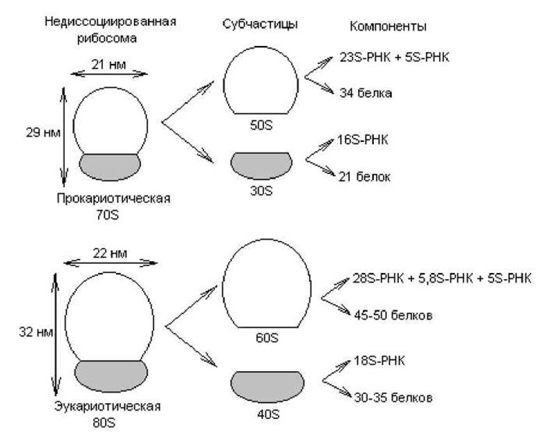
Fig. 3.12. Structure and composition of prokaryotic and eukaryotic ribosomes
Electron Cell/15.html">Microscopy studies of 70S ribosomes have shown that the small and large subunits contact each other at several points, forming a cleft between them that is necessary to accommodate the mRNA during translation. Two functionally important sites on the 70S ribosome are crucial for understanding the translation process: the A site (aminoacyl site), which serves for the binding of aminoacyl-tRNA, and the P site (peptidyl site), which binds the growing peptide chain.
In addition to aminoacyl-tRNAs and ribosomes, translation involves A large number of accessory proteins known as initiation, elongation, and METABOLISM/31.html">Transcription termination factors.
The Essence of translation lies in the sequential decoding of mRNA in the 5' → 3' direction with the aid of aminoacyl-tRNAs, accompanied by the successive Condensation of amino acid residues, starting from the amino (N)-terminal end of the polypeptide chain toward the carboxyl (C)-terminal end. The template principle of the process is maintained through the recognition of complementary NUCLEOTIDES between the successive mRNA codon and the tRNA anticodon. Translation has been studied most thoroughly in prokaryotes, and The Mechanism of this process will be examined using E. coli translation as a model.
Translation initiation. Reading of the mRNA begins with the AUG codon, which marks the 5' end of the coding sequence and specifies the N-terminal (first) amino acid of the synthesized polypeptide. Initiation requires the 30S ribosomal subunit, which forms a complex with initiation factors (IF1, IF2, IF3), GTP, and fMet-tRNA. This complete complex binds to the 5' end of the mRNA coding sequence near the AUG codon. Apparently, IF2 is able to distinguish fMet-tRNA (formylmethionine-tRNA) from regular Met-tRNA, which binds to internal AUG codons within the mRNA but cannot initiate translation from the start AUG codon. This specificity is conferred by the N-formyl group, which is absent in Met-tRNA.
The start codon is recognized in the following manner. The binding of the 30S subunit to mRNA is strictly controlled by a nucleotide sequence located approximately 10 nucleotides upstream of the 5' end of the start codon. This interaction is facilitated by complementary base-pairing between this purine-rich sequence and a polypyrimidine stretch within the 16S rRNA. The initiation process depends on various Structural Features of the interacting regions, including the Introduction/11.html">Secondary structure of the mRNA segment containing the start AUG codon. This plays an important role in regulating the efficiency of protein synthesis.
Thus, during initiation, this complex binds to the P site of the 30S ribosomal subunit, with formylmethionine serving as the first amino acid in the peptide. This is followed by the attachment of the 50S ribosomal subunit to form the 70S initiation complex (Fig. 3.13). The energy for initiating Protein synthesis is provided by the hydrolysis of GTP to GDP and Pi.
Translation elongation. For the Formation of the first peptide bond, the aminoacyl-tRNA corresponding to the next codon must occupy the A site of the ribosome. To achieve this, the aminoacyl-tRNA must first bind the elongation factor EF-Tu and GTP. The resulting ternary complex (aminoacyl-tRNA–EF-Tu–GTP) delivers the aminoacyl-tRNA to the A site. At this point, GTP is hydrolyzed, and the EF-Tu–GDP complex is released from the ribosome. Once both the A and P sites are occupied, the peptidyl transferase activity of the 50S subunit catalyzes the transfer of the fMet group from its tRNA to the amino group of the aminoacyl-tRNA located in the A site (Fig. 3.14). As a result, a dipeptidyl-tRNA occupies the A site, while a deacylated tRNA resides in the P site (Fig. 3.13).
The peptidyl transferase activity of ribosomes appears to be associated not with the protein moiety of the 50S subunit, but with one of its RNA components—a ribozyme.
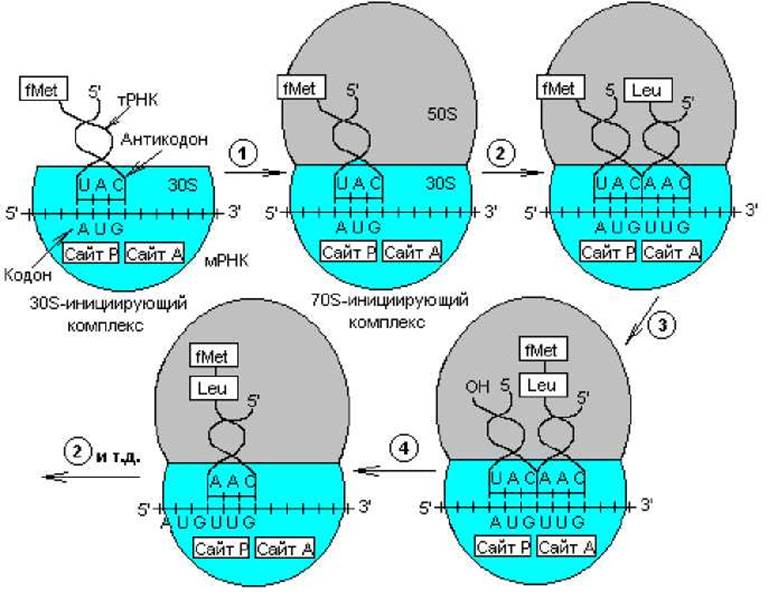
Fig. 3.13. Translation of the Genetic Code: 1 — formation of the 70S initiation complex; 2 — binding of aminoacyl-tRNA to the ribosomal A site; 3 — peptide bond formation; 4 — ribosomal translocation. During elongation, steps 2–4 are repeated
To read the next codon and extend the polypeptide chain by one more amino acid, the entire series of reactions must repeat. Before this happens, however, the uncharged tRNA vacates the P-site, the newly formed peptidyl-tRNA shifts to it from the A-site (without codon-anticodon interaction), and the ribosome moves forward in a stepwise manner (by 3 nucleotides) toward the 3'-end of the mRNA. All these processes are driven by the elongation factor EF-G during GTP-dependent ribosomal translocation. As a result of these three events, the A-site is cleared and the next codon is exposed, allowing the subsequent elongation cycle to begin (Fig. 3.13). It should be noted that the formation of each peptide bond consumes an energy equivalent to four energy equivalents (assuming the energy of a phosphate bond formation as one equivalent): two ATP equivalents are utilized during tRNA aminoacylation and two GTP equivalents in each elongation cycle.

Fig. 3.14. Formation of the peptide bond between the first Two amino acids on ribosomes
Translation termination. The process of sequential codon translation eventually reaches a point where one of the three stop codons—UAG, UAA, or UGA—enters the A-site. In nature, there are no tRNAs with anticodons matching these codons. Here, termination factors RF-1 and RF-2 come into play, catalyzing the release of the polypeptide chain from the tRNA, the release of the tRNA from the ribosome, and the dissociation of the 70S ribosome from the mRNA.
Following translation initiation, the 70S ribosome moves away from the initiation site as each successive codon is read. Once the distance between the ribosome and the initiation site reaches 100–200 nucleotides, a new initiation event can occur at that site. Furthermore, as soon as the second ribosome covers the same distance, a third initiation can take place, and so on. Thus, a single protein-coding mRNA sequence can be translated simultaneously by multiple ribosomes. Such multiribosomal translation complexes are called polyribosomes or Polysomes.
Messenger RNAs consisting of multiple protein-coding regions are often translated sequentially: when a ribosome reaches the stop codon of the first sequence, it dissociates from the mRNA, and a new complex binds to the next initiation site. Sometimes this does not happen, and the ribosome translating the first coding sequence continues to move along the mRNA without detaching, initiating translation at other sites.
In some cases, the translation of the first coding sequence can begin and even finish before the Transcription of the remaining sequences is complete, as is the case, for example, with the lac or trp operons of E. coli.
Features of translation in eukaryotes. The translation of eukaryotic mRNA is generally similar to that of prokaryotes. However, there are several key differences. First, transcription and translation machinery in eukaryotes are separated in both time and space, since transcription occurs in The Nucleus, whereas translation takes place in the Cytoplasm. Second, the initiating aminoacyl-tRNA in eukaryotes is not fMet-tRNA, but a specialized initiator Met-tRNA. Third, the 5' and 3' ends of eukaryotic mRNAs feature specific structures—caps and tails—that take part in translation. It is known that certain translation initiation factors recognize the capped regions to bind to the mRNA and initiate the translation process.
Last update: 06/08/2026
Editorial and Educational Adaptation: This material has been compiled based on the primary/original source text. The project team performed an editorial review, corrected technical inaccuracies, structured sections, and adapted the content for an educational format.
What was processed:
- elimination of formatting defects (OCR errors, structural breaks, corrupted characters);
- editorial organization of content;
- standardization of terminology in accordance with academic sources;
- verification of factual statements against the original source text.
All mentions of the author, publication year, and origin of the primary text have been preserved in accordance with the source.