Practical Protein Chemistry - A. Darbre 1989
Traditional strategy for protein structure determination
Choosing a research strategy
Figure 10.2 illustrates the strategic workflow for Cell/13.html">Protein Structure analysis in single- and two-step approaches. The starting protein sample must be purified to homogeneity (at least 95% purity), and Lipids, CARBOHYDRATES, and Cofactors must be removed.
The choice of Proteolytic Enzymes for initial fragmentation (in the single-step approach) is determined by the molecular weight and Amino Acid Composition of the protein, as these parameters allow for predicting the average size of the Peptides generated upon Hydrolysis (e.g., by Trypsin at Lysine and Arginine residues). Typically, the most specific enzymes are employed—trypsin, Chymotrypsin, staphylococcal protease, and the protease from A. mellea (Table 10.1). To avoid the difficulties associated with separating the hydrolysate of a large protein that contains a significant number (>40) of peptides, certain potential Cleavage sites in the polypeptide chain can be blocked, or a two-step analysis scheme can be used. For instance, tryptic cleavage at lysine residues can be prevented by modifying them with maleic anhydride [8]. Following tryptic hydrolysis, the modified fragments are unblocked by acid Treatment prior to Separation (via Ion-exchange Chromatography or Electrophoresis). Similarly, peptide bonds formed by arginine residues can be protected from trypsin action by pre-treating the protein with cyclohexanedione in the presence of borate [48]. A prerequisite for the successful application of this approach is achieving complete modification and subsequent removal of the protecting groups. The blocking technique, combined with diagonal electrophoresis Methods [8, 48, 49], is used for the selective isolation of tryptic peptides containing internal lysine or arginine residues. Alternatively, primary fragmentation of the polypeptide chain exclusively at arginine or lysine residues can be achieved using specific enzymes: Clostripain [43] and the A. mellea protease [65], respectively.
In the two-step analysis scheme, large protein fragments are generated either by chemical cleavage methods (Table 10.2) targeting Amino Acids that occur relatively rarely in the polypeptide chain—such as Tryptophan, Methionine, and Cysteine—or by Limited proteolysis of the native protein [36, 40, 51, 52]. Subsequent fragmentation of each resulting large peptide yields significantly fewer components than fragmentation of the whole protein molecule, which greatly simplifies further fractionation. However, achieving complete and specific chemical Cleavage of the protein, successfully separating the large fragments, and ultimately determining their order within the chain can present considerable challenges. For example, Cyanogen bromide cleaves the polypeptide chain at methionine residues, converting them into homoserine residues; however, the oxidized form of methionine—methionine sulfoxide—is resistant to the reagent, and the Met–Ser and Met–Thr bonds are cleaved only partially. Under conditions of acid hydrolysis of labile Asp–Pro bonds, tryptophan undergoes oxidative degradation, and the amides of dicarboxylic Amino acids are partially destroyed. The large denatured Polypeptides produced by chemical methods are often poorly soluble and fail to elute from ion-exchange columns. The Diversity of charged forms for each peptide, resulting from partial deamidation or The formation of mixtures of peptides with C-terminal homoserine and homoserine lactone residues, further complicates their charge-based separation. Reconstructing the polypeptide chain from cyanogen bromide fragments requires the isolation of all methionine-containing peptides from a separate hydrolysate of the original protein.
Class="center">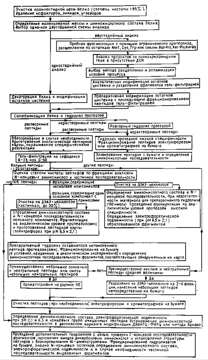
FIG. 10.2. Strategy for amino acid Sequence Determination (non-automated methods).
Table 10.1. Specificity of Proteolytic Enzymes
|
Enzyme |
Residues X at which protease action predominantly occurs |
Remarks |
|
Trypsin |
Basic amino acid residues: C-terminal Lys, Arg, and aminoethyl-Cys [67] |
Cleavage does not occur at X–Pro bonds; less efficient if an acidic residue precedes or follows X; more specific for Arg at alkaline pH values [22]; proceeds in 2 M urea |
|
Chymotrypsin |
Aromatic amino acid residues: C-terminal Trp, Tyr, Phe, Leu [67] |
Cleavage does not occur at X–Pro bonds and oxidized Trp; less efficient if an acidic residue precedes or follows X; proceeds in 2 M urea |
|
Aliphatic amino acid residues: Ala, Val, Gly, Ser [27] |
Under partial Digestion conditions, cleavage sites are unpredictable; cleavage proceeds in 2 M urea |
|
|
Staphylococcal protease |
Acidic amino acid residues: C-terminal Glu and Asp [15, 16, 32] |
Conditions can be adjusted for cleavage exclusively at Glu or at both Glu and Asp; cleavage also occurs at carboxymethyl-Cys; less efficient if X is followed by a residue with a branched side chain or if X is surrounded by acidic residues; proceeds in 4 M urea [15] |
|
N-terminal Ile, Leu, Val, Phe [29, 41, 42] |
Cleavage does not occur at X–Pro bonds; proceeds at 60–80 °C, which can facilitate peptide dissolution |
|
|
Aromatic amino acid residues: N-terminal Trp, Tyr, Phe, Leu [67] |
Also cleaves many other bonds; acidic hydrolysis conditions may assist in peptide dissolution |
|
|
Protease from Armillaria mellea |
N-terminal Lys and aminoethyl-Cys [65] |
Also cleaves at other residues, such as N-terminal Arg, Phe, Trp (G. Weber, unpublished data) and C-terminal Arg [13] |
Table 10.2. Chemical Cleavage Methods Used in the Two-Step Analysis Scheme
|
Reagent |
Primary specificity |
Partial cleavage |
|
|
Cyanogen bromide [23] |
At Met, yielding C-terminal homoserine and homoserine lactone residues; the latter is resistant to Carboxypeptidases |
Met-oxide bond is not cleaved; Met–Ser, Met–Thr [62], Met–(Cys–Cys) [14], and Met–Glu [10] bonds are partially cleaved |
Acid cleavage at Asp–Pro and Asn–Pro bonds [50] and oxidative cleavage at Trp [3] |
|
2-Nitro-5-thiocyanobenzoic acid followed by alkaline cleavage [11] |
At Cys, yielding an N-terminal 4-carboxyiminothiazolidine that blocks the Edman reaction and aminopeptidase activity; deblocking is possible via catalytic reduction [57] |
Formation of mixed disulfides [54] and β-elimination of the S-cyano derivative [70] |
Phe–Thr and Phe–Ser bonds when catalytic reduction is used to deblock the N-terminal residue [57] |
|
Dimethyl sulfoxide hydrobromide [56] or N-chlorosuccinimide [64] |
At Trp, yielding a C-terminal lactone; Cys and Met are oxidized |
||
|
BrCN in formic and heptafluorobutyric acid [47] |
At Trp, provided Met is pre-photoxidized |
||
|
Hydroxylamine [5] |
At Asn–Gly |
Extent of cleavage depends on prior exposure of the peptide to acidic or alkaline conditions [38] |
Asn–Leu [6] and Asn–Ala [71] bonds |
|
Dilute acid [50] |
At Asp–Pro and Asn–Pro |
Many of these complications can be proactively avoided. For instance, prior to hydrolysis, methionine sulfoxide residues can be reduced back to methionine by treating the protein with 2-mercaptoethanol or dithiothreitol under denaturing conditions. It should be noted that methionine sulfoxide is not detected during Amino acid analysis of a protein because it is reduced to methionine under acidic hydrolysis conditions; however, it can be quantified using indirect methods [63]. The peptide mixture containing C-terminal homoserine and homoserine lactone residues can be converted entirely into either of these forms [2, 45].
For dissolving and separating large peptides, 8 M urea followed by ion-exchange chromatography in the same solution [25] can be used, or 100% formic acid coupled with Gel filtration on Bio-Gels in 20–70% formic acid [1]. Solubilization can also be achieved by modifying the lysine residues of the peptides with citraconic anhydride; fractionation is then carried out on Bio-Gel or Sephadex columns at neutral pH.
In the final stages of analysis, a targeted search can be conducted for peptides required to link large fragments—the so-called "overlapping" peptides. For example, methionine-containing peptides can be selectively isolated using diagonal electrophoresis [73]. The optimal method for identifying cysteine-containing peptides involves pre-labeling cysteine residues with [14C]iodoacetic acid followed by the targeted isolation of radioactive fragments [78, 79]. Tryptophan-containing peptides are detected in chromatographic peaks by their maximal absorbance at A280 and characteristic fluorescence or specific staining during paper chromatography.
Since gel filtration unquestionably yields better resolution in the separation of large peptides than ion-exchange chromatography, the optimal Protein Cleavage method for a two-step analysis (Fig. 10.2) is best selected based on the results of analytical Polyacrylamide gel electrophoresis (PAGE) in the presence of sodium dodecyl sulfate (SDS). When protein bands are clearly resolved in the gel, gel filtration is the preferred method for the preparative fractionation of peptides. SDS-PAGE is also employed to monitor the completeness of chemical cleavage reactions on the protein.
Last update: 06/08/2026
Editorial and Educational Adaptation: This material has been compiled based on the primary/original source text. The project team performed an editorial review, corrected technical inaccuracies, structured sections, and adapted the content for an educational format.
What was processed:
- elimination of formatting defects (OCR errors, structural breaks, corrupted characters);
- editorial organization of content;
- standardization of terminology in accordance with academic sources;
- verification of factual statements against the original source text.
All mentions of the author, publication year, and origin of the primary text have been preserved in accordance with the source.