Molecular Biology: Protein Structure and Function - Stepanov V.M. 2005
Quaternary protein structure
Stoichiometry and geometry of quaternary structure
The subunits that form The quaternary Structure of a protein can vary significantly both in their architecture and functional properties. Such Proteins are classified as heteromeric. They occur quite frequently, making it possible to integrate several interrelated Functions into a single structure and create a multifunctional molecule. Protein kinase is a classic example of a heteromer, where one subunit (C, the catalytic subunit) is responsible for the actual enzymatic activity and catalyzes The transfer of a phosphate group from ATP to a protein, while the other is regulatory (R). In the absence of cyclic AMP, the latter binds to the C-subunit to form the R—C complex and inhibits it. Upon forming a complex with cAMP, the quaternary structure dissociates, and the C-subunit becomes capable of phosphorylating protein substrates. RNA polymerase serves as another striking example of a heteromeric protein.
In homomeric proteins, the subunits are identical. Similar to them are heteromeric proteins which, strictly speaking, consist of non-identical subunits, yet are quite similar both in their polypeptide chain folding pattern and function.
Proteins composed of two, three, four, and so on, subunits occur with vastly differing frequencies. For instance, in a more or less random sample of approximately 200 proteins with a Molecular Weight of no more than 300 kDa, the distribution was as follows: 102 dimers, 53 tetramers, 23 hexamers, whereas there were only 9 trimers, no pentamers, and 3 octamers. Thus, the overwhelming majority of proteins possessing a quaternary structure are dimers, tetramers, and hexamers, with the latter being found in proteins with a molecular weight exceeding 100 kDa.
The geometry of symmetrical dimers is straightforward. The same applies to trimers, which are quite rare yet do occur. In particular, they can play a vital role in The formation of transmembrane channels, as a sort of bundle formed by three subunits naturally creates an internal pore. In tetrameric proteins, subunits may be positioned at the corners of a square—which is rare—or occupy the vertices of a tetrahedron (Fig. 7.2). This latter type of quaternary structure, in which each subunit interacts with the remaining three with varying degrees of strength while the molecule as a whole remains highly compact, is observed with notable frequency. Hexameric proteins are characterized by
an octahedral packing, whereas flat hexagonal structures are much less common.
The subunits that form a symmetrical quaternary structure may be identical, but they can also differ while remaining of the same type—evolutionarily related proteins that share the same pathway of spatial folding of the peptide chain. In such cases, the Formation of the quaternary structure exhibits distinct features. If, for instance, a tetrameric protein is formed by structurally homologous, similar a and ß subunits, two situations are possible depending on the degree of their divergence. In the first case, when the a—a, ß—a, and ß—ß contacts differ significantly, a Quaternary Structure of the a2/ß2 type is formed, while other subunit combinations remain unstable and are practically undetectable. This is precisely how Hemoglobin molecules are structured. However, if the intersubunit contacts are practically equivalent, a full set of possible quaternary structures can form—namely, a4, a3 / ß1, a2 / ß2 > a1 / ß3, and ß4—with the proportion of each of these multiple forms (note that this is far from the only cause for the appearance of multiple protein forms) determined by the relative Abundance of the a and ß subunits.
Class="center">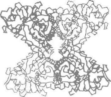
Fig. 7.2. Quaternary structure of rabbit Skeletal Muscle aldolase.
The enzyme consists of four identical subunits located at the vertices of a tetrahedron (two, highlighted by bold lines, are positioned closer to the reader). In this case, intersubunit interactions are maintained through hydrophobic contacts between the side chains of Amino Acids that form a-helices
A typical example is human Lactate dehydrogenase, which is built from four subunits. These can be of two types: the M-subunit (from muscle), which predominates in smooth muscle, and the H-subunit (from Heart), which is predominantly synthesized in cardiac muscle. For lactate dehydrogenase, the following set of multiple forms is possible: M4, M3H1, M2H2, M1H3, and H4. It should be noted that multiple forms whose differences are genetically determined rather than caused by post-translational modifications or protein damage are conventionally called isoforms. The difference in the Introduction/19.html">Primary Structure of the subunits affects The ratio of cationic and anionic groups, leading to differences in the charge of the isoforms and allowing them to be easily separated by Electrophoresis. Such an Analysis of the isoenzyme composition of Blood lactate dehydrogenase makes it possible to monitor the release of lactate dehydrogenase from certain Organs—particularly The Heart—into the bloodstream during The Development of corresponding pathologies, such as myocardial necrosis. In the latter case, the content of isoforms enriched in the H-subunit increases. This approach has found widespread application in medical Diagnostics and biochemical genetics.
Last update: 13/08/2026
Editorial and Educational Adaptation: This material has been compiled based on the primary/original source text. The project team performed an editorial review, corrected technical inaccuracies, structured sections, and adapted the content for an educational format.
What was processed:
- elimination of formatting defects (OCR errors, structural breaks, corrupted characters);
- editorial organization of content;
- standardization of terminology in accordance with academic sources;
- verification of factual statements against the original source text.
All mentions of the author, publication year, and origin of the primary text have been preserved in accordance with the source.