Molecular Biology: Protein Structure and Functions - Stepanov V.M. 2005
Quaternary protein structure
Quaternary Structure refers to the spatial arrangement of interacting subunits formed by individual protein polypeptide chains.
The formation of quaternary structure involves not the peptide chains themselves, but rather the globules formed by each of these chains independently. Thus, METABOLISM/2.html">THE CONCEPT OF quaternary structure refers to an ensemble of globules. The interaction between the latter is strong enough for the ensemble to act as a single molecule; at the same time, each associated globule—or subunit—retains a significant degree of autonomy, which is typically much more pronounced than the autonomy of a domain within a tertiary structure.
However, cases are known where two or more Polypeptides make up a single globule. As a rule, this is the result of Limited proteolysis—the localized Cleavage of an originally intact polypeptide chain (which formed a globule According to the standard rules of tertiary structure formation) into separate segments. Naturally, such Proteins should not be classified as having a quaternary structure.
Insulin serves as a clear example. Its monomer is built from two peptide chains, A and B, containing 21 and 30 amino acid residues, respectively. However, these chains form a unified structure produced after the connecting peptide C is excised from the proinsulin molecule, so there are no grounds to consider them subunits. Another example is concanavalin A (mol. mass 26 kDa). This plant protein belongs to the Lectins and specifically binds to carbohydrate components on The Cell surface. Purified concanavalin is a mixture of intact and proteolysis-damaged molecules, with the peptide chain in the latter being cleaved into two fragments with molecular weights of 20 and 6 kDa. Clearly, such damaged molecules do not differ fundamentally in their structural Organization from intact ones, and there is no reason to ascribe a quaternary structure to them despite the presence of two peptide chains.
It should be emphasized once again that we are discussing proteins that form a single globule. In some proteins, the polypeptide chain forms multiple globules (domains) upon folding, between which a system of non-covalent interactions is established. Subsequent proteolytic Cleavage of the chain regions connecting the domains makes them fully autonomous, elevating them to the rank of subunits that form a quaternary structure.
The term "aggregate" is sometimes used in the literature as a synonym for "quaternary structure," which is difficult to justify since the latter implies a very high level of organization—the assembly of subunits into a molecule stabilized by a system of non-covalent interactions. Likewise, there is no justification for classifying supramolecular (e.g., multienzyme) complexes or extended structures, such as phage coats (Fig. 7.1) or protein crystalline inclusion bodies in certain bacilli, as quaternary structures, although the mechanisms of their formation share much in common with quaternary structure assembly.
Quaternary structure represents the final level of protein molecular organization, and an optional one at that: up to half of known proteins lack it. The boundary between proteins that possess a Quaternary Structure and those that do not is not always entirely sharp. Some proteins dissociate into subunits relatively easily even under physiological conditions. At the same time, the formation of more or less stable dimers is known in solutions of certain proteins that are not classified as having a quaternary structure—pancreatic Ribonuclease is a case in point. Note that protein surfaces, densely packed with functional groups and highly irregular, tend to form at least a weak system of interactions, which underlies protein crystallization, among other things. Such interactions should seemingly be viewed as a kind of precursor that could serve as an evolutionary starting point for The Development of a sufficiently refined quaternary structure.
Class="center">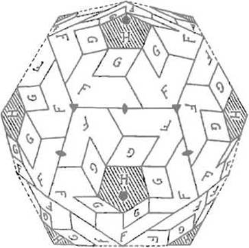
Fig. 7.1. Arrangement of proteins (F, G, H) forming the capsid coat of phage φX174.
Filled pentagons denote fivefold axes of Symmetry, triangles denote threefold axes, and lens-shaped symbols denote twofold axes
Last update: 13/08/2026
Editorial and Educational Adaptation: This material has been compiled based on the primary/original source text. The project team performed an editorial review, corrected technical inaccuracies, structured sections, and adapted the content for an educational format.
What was processed:
- elimination of formatting defects (OCR errors, structural breaks, corrupted characters);
- editorial organization of content;
- standardization of terminology in accordance with academic sources;
- verification of factual statements against the original source text.
All mentions of the author, publication year, and origin of the primary text have been preserved in accordance with the source.