MICROBIOLOGY Study Guide - 2012
CHAPTER 5. FUNGI
5.2. CLASSIFICATION OF FILAMENTOUS FUNGI. CHARACTERISTICS OF SELECTED REPRESENTATIVES
Currently, Fungi are classified under the superkingdom Eukaryota and the kingdom Mycota (Fungi), which comprises two phyla: slime Molds (mycota) and true fungi (Eumycota). The phylum Eumycota is divided into seven classes based on their Structure and developmental stages, specifically the anamorph (asexual) and teleomorph (sexual) stages. Four classes belong to lower fungi: Chytridiomycetes, Hyphochytridiomycetes, Oomycetes, and Zygomycetes, while three belong to higher fungi: Ascomycetes, Basidiomycetes, and Deuteromycetes.
Lower fungi possess two distinct features that differentiate them from other fungi. First, they feature a unicellular mycelium that lacks cross-walls, known as a aseptate mycelium (from the Greek septum meaning partition). Second, the spores of lower fungi are always formed endogenously, inside specialized Cells called sporangia. The defining characteristics of higher fungi include a multicellular septate mycelium and exogenous spores called conidia.
1. Class Chytridiomycetes — chytrids. These are predominantly aquatic fungi, although some species are found in soil. They possess a rudimentary mycelium (or lack it entirely), and their body consists of a naked protoplast that becomes enclosed in a Cell wall upon transforming into a sporangium. Chytrids reproduce mainly asexually via motile zoospores that develop within sporangia. Parasitic fungi in this group include Synchytrium endobioticum, the CAUSATIVE AGENT OF potato wart disease.
2. Class Hyphochytridiomycetes — hyphochytrids, a unicellular form of fungi that lack a sexual reproduction stage. Most species in this class are intracellular parasites of green and brown Algae, as well as cultivated plants. In particular, Plasmodiophora brassicae is the causative agent of clubroot in cruciferous crops, while Spongospora subterranea causes powdery scab in potatoes.
3. Class Oomycetes — oomycetes. This class includes both aquatic and terrestrial forms that reproduce both asexually via zoospores and sexually through The formation of oospore. Oomycetes include the phytopathogenic species Phytophthora infestans, the causative agent of late blight in potatoes, tomatoes, and eggplants. Short, branched sporangiophores bear egg-shaped or lemon-shaped sporangia. When sporangia come into contact with wet soil, they produce zoospores, which then germinate into hyphae. Another notable oomycete is Plasmopara viticola, the causative agent of downy mildew in grapes.
4. Class Zygomycetes — zygomycetes. This is the most highly evolved group of lower fungi that has transitioned to a terrestrial lifestyle. Among zygomycetes, the most widespread species are described below.
Mucor mucedo — pin mold or black bread mold. It is found on decaying organic substrates or food products as a fluffy white or grey felt-like coating. When reproducing, Mucor forms a heavily branched, single-celled mycelium. Unicellular spore-bearing hyphae, known as sporangiophores, arise from the substrate mycelium and terminate in a large spherical sporangium (from the Greek spora meaning seed and angion meaning vessel) (Fig. 12; 1).
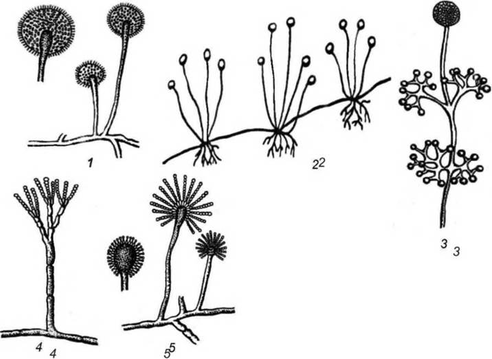
Fig. 12. Structure of selected representatives of filamentous fungi:
1 — Mucor, 2 — Rhizopus; 3 — Thamnidium, 4 — Penicillium, 5 — Aspergillus
Rhizopus nigricans causes tissue destruction in various fruits, vegetables, and ROOT crops, leading to spoilage known as "soft rot". Fungi of the genus Rhizopus rapidly spread across The surface of a substrate by sending out horizontal hyphae in all directions, known as stolons—runners resembling strawberry runners. These runners anchor to the substrate using root-like structures called rhizoids. Bundles of sporangiophores rise upward from the rhizoids. The unbranched sporangiophore expands at the apex to form a rounded sporangium, which is separated by a septum from the fertile hypha. A large number of spherical spores develop inside the sporangium. Upon rupture of the sporangium, the asexual sporangiospores are released and dispersed by air currents. Upon landing on a suitable substrate, they germinate to form a new vegetative mycelium (Fig. 12; 2).
Thamnidium elegans features sporangiophores with Two Types of sporangia: a large, multi-spored sporangium at the apex, and smaller sporangia (sporangioles) containing a limited number of spores on the lateral Branches of the sporangiophore (Fig. 12; 3).
Fungi of the genus Thamnidium are cold-tolerant and frequently cause food spoilage during refrigerated storage.
5. Class Ascomycetes — ascomycetes. This class comprises higher fungi with a well-developed multicellular mycelium. The primary spore-bearing organ in ascomycetes is the sac or ascus (from the Greek askos meaning bag), which gives this class of fungi its name. The ascus stage represents the final phase of the Sexual process in ascomycetes. Many ascomycetes can reproduce asexually through exospores called conidia. This form of sporulation is referred to as the imperfect state. The most common representatives of ascomycetes are fungi of the genera Penicillium and Aspergillus.
The genus Penicillium possesses a branched mycelium that gives rise to septate spore-bearing hyphae known as conidiophores. At the tips of the conidiophores are cellular outgrowths called sterigmata, arranged in a brush-like manner, which is why penicilla are referred to as brush molds. The terminal sterigmata end in chains of conidia (Fig. 12; 4). In different species of Penicillium, the conidia vary in color—green, blue, or grey-green—which determines the coloration of the mold colony.
Penicilla frequently proliferate on food products such as grain, bread, pasta, meat and dairy products, fruits, and vegetables. In Fermentation industries, this fungus is found on barley and malt. Conidia of Penicillium are abundant in the air.
Certain species of the genus Penicillium produce the antibiotic penicillin (Pen. notatum, Pen. chrysogenum). In the dairy industry, mold fungi of the species Pen. roqueforti and Pen. camemberti are used in The production of cheeses such as Roquefort and Camembert.
Genus Aspergillus. Unlike other higher fungi, fungi of the genus Aspergillus possess aseptate conidiophores. The tips of the conidiophores expand into a clavate (club-shaped) Swelling, from the surface of which numerous short sterigmata with chains of conidiospores radiate in all directions. The conidia are arranged radially, resembling Water jets spraying from a watering can, which gave rise to the alternative name for aspergilli—water-mold or duster mold (Fig. 12; 5). Aspergilli conidia exhibit various colors: green, yellow, brown, and black. A widely distributed species is Aspergillus glaucus, which is characterized by green or grey-green colonies and frequently grows on bread, jam, preserves, and marmalade.
Certain Representatives of the genus Aspergillus are used for the industrial production of citric acid, oxalic acid, and other organic acids (Aspergillus niger), as well as enzyme preparations such as amylase and avamorin (A. oryzae, A. awamori). Some aspergilli produce Antibiotics, while others include human pathogens.
Claviceps purpurea is a phytopathogenic fungus that causes ergot in rye. The ascospores of the fungus infect the Ovaries of rye during flowering. As the fungus develops within the cereal spikes, dark purple, hard horns known as sclerotia appear in place of the grains. Sclerotia contain potent Alkaloids (lysergic acid derivatives) that are toxic to humans and animals.
6. Class Basidiomycetes — basidiomycetes. This is a vast group of highly evolved multicellular fungi. The primary spore-bearing structure of basidiomycetes is the basidium. A distinction is made between fungi with unicellular basidia, which bear two to four basidiospores on short outgrowths (sterigmata), and fungi with multicellular basidia, consisting of four elongated cells, each supporting a single basidiospore on a sterigma.
Basidiomycetes with unicellular basidia include:
✵ cap fungi (tubular, gilled, and tooth fungi) that feature a single-year fruiting body shaped like a cap attached to a stalk;
✵ bracket fungi (polypores) that grow on tree trunks and destroy wood;
✵ dry rot fungi that multiply on the wooden Structural elements of buildings. They can also be found in wine cellars on barrels, shelves, and other wooden items. As these fungi develop, the wood gradually breaks down and decays.
Many parasitic fungi that affect field, vegetable, and garden crops belong to the basidiomycetes. Their most prominent representatives are smut fungi and rust fungi.
Smut fungi primarily attack cereal crops, causing a disease known as smut. Plants infected by the fungus appear charred, which is how the pathogen got its name.
Based on how their chlamydospores germinate, smut fungi are divided into two families: Ustilaginaceae (represented by U. tritici) and Tilletiaceae (represented by Т. avenae). Germinated chlamydospores of Ustilaginaceae form a four-celled basidium, whereas those of Tilletiaceae form a single-celled basidium.
Rust fungi (genus Puccinia) are pathogenic to many cultivated plants. These fungi derived their name from the characteristic rust-colored spots that appear on the infected PARTS OF THE plants.
7. Class Deuteromycetes (imperfect fungi). This class includes all fungi whose Classification into other groups has not been established and whose sexual reproduction cycle has not been identified. Most imperfect fungi reproduce via conidia, which vary in shape, structure, arrangement, and color. This group of fungi is heterogeneous, as there are quite a few imperfect forms among ascomycetes, basidiomycetes, and others.
Deuteromycetes are widespread in nature, and many of them are responsible for the spoilage of fruits, vegetables, and foodstuffs. Among them are species that cause diseases in humans, animals, and plants. The following fungi are most frequently responsible for food spoilage.
Genus Fusarium. The mycelium of the fungus can be white, pink, or lilac. Fusarium produces two types of conidia: sickle-shaped, multicellular macroconidia and smaller, elliptical or rounded, unicellular microconidia (Fig. 13; 1). Certain species of the genus Fusarium cause fruit and vegetable diseases collectively known as "fusariosis" (see Section 15.12).
Genus Botritis. The arborescent conidiophores of this fungus bear smoke-colored, unicellular conidia clustered in heads at the branch tips. Small, black sclerotia frequently appear on the mycelium (Fig. 13; 2). Botrytis affects apples, pears, and many berries.
Genus Phoma. The fungus features short conidiophores within pycnidia, producing colorless, elliptical conidia. Representatives of this genus cause a disease in vegetable crops known as phoma rot (Fig. 13; 3).
Genus Catenularia (Fig. 13; 4) forms small, slow-growing colonies of a chocolate-brown color. The conidia are shiny and yellow-brown, extending from the terminal hyphae of the aerial mycelium in long chains. Catenularia fuliginea frequently multiplies in sweetened condensed milk, forming chocolate-colored clumps or "buttons."
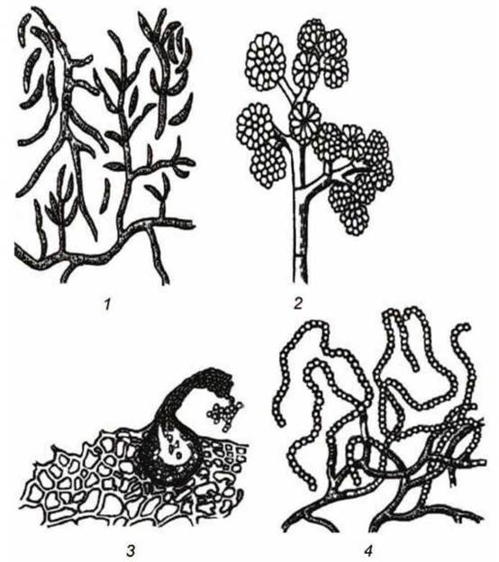
Fig. 13. Structure of mycelial fungi of the class Deuteromycetes:
1 — Fusariunr, 2 — Botritis; 3 — Phoma; 4 — Catenularia
Genus Cladosporium. The fungus possesses sparsely branched conidiophores that terminate in chains of conidia of various shapes: round, oval, or cylindrical (Fig. 14; 1). Developing on foodstuffs (butter, cheese, fruits, etc.), Cladosporium forms velvety, olive-green colonies on them.
These fungi secrete a black pigment into the surrounding medium, causing the surface of the infected products to gradually darken.
Endomyces lactis — synonym Geotrichum candidum — dairy mold (Fig. 14; 2). A white, velvety bloom of dairy mold frequently forms On the surface of dairy products during prolonged storage. It possesses a heavily branched mycelium whose hyphae break apart into individual cells known as oidia or arthrospores. Dairy mold contains active Enzymes that break down Proteins and fats.
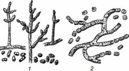
Fig. 14. Mycelial deuteromycete fungi:
1 — Cladosporiunr, 2 — Endomyces lactis
Last update: 13/08/2026
Editorial and Educational Adaptation: This material has been compiled based on the primary/original source text. The project team performed an editorial review, corrected technical inaccuracies, structured sections, and adapted the content for an educational format.
What was processed:
- elimination of formatting defects (OCR errors, structural breaks, corrupted characters);
- editorial organization of content;
- standardization of terminology in accordance with academic sources;
- verification of factual statements against the original source text.
All mentions of the author, publication year, and origin of the primary text have been preserved in accordance with the source.