Fundamentals of Molecular Biology - V.I. Rezyapkin 2009
Genome Organization
Replication of Genomes in DNA-Containing Viruses
A large number of Proteins are involved in the Replication of DNA virus genomes, including DNA polymerases, primases, ligases, helicases, replication initiation proteins, etc. Some of these are Enzymes, while others are not. These Proteins can be of either viral or cellular origin. For instance, in phage φX174, all proteins participating in replication—with the exception of protein A—are of cellular origin, whereas in phage T-4, they are of viral origin. There are also instances where replication proteins are of mixed origin, with some subunits encoded in the cellular genome and others in the viral genome.
Adenovirus Genome Replication
The Role of the primer in adenovirus DNA genome replication is performed by a complex consisting of a virus-specific terminal protein and a single covalently attached nucleotide (Fig. 11.2). Following the interaction of this complex, Synthesis of the daughter strand commences. During this process, the parental strand is displaced from the duplex. A nucleotide-protein primer attaches to the 3’ end of the released strand, and synthesis of the complementary strand begins. As a result, a double-stranded DNA molecule is formed.
Class="center">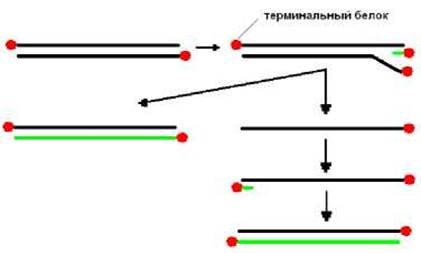
Fig. 11.2. Schematic of adenovirus replication. SV40 genome replication
The SV40 genome is represented by a double-stranded, circular, covalently closed DNA molecule.
Upon interaction of the virus-specific helicase (T-antigen) with THE ORIGIN OF replication, the primase generates primers on both parental strands. Subsequently, daughter strand synthesis proceeds. The torsional stress generated by DNA unwinding is relieved by topoisomerase I. Replication yields two linked DNA molecules, which are then segregated with the participation of topoisomerase II (Fig. 11.3). All proteins involved in Viral METABOLISM/36.html">DNA replication, except for the T-antigen, are of cellular origin.
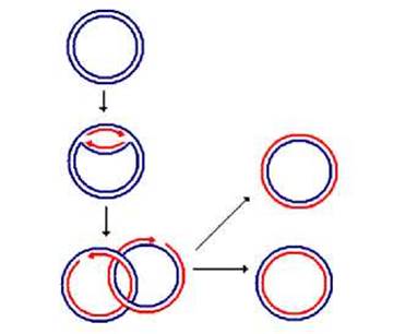
Fig. 11.3. Schematic of SV40 genome replication
Replication of Phage λ
The genomic nucleic acid of phage λ is a linear double-stranded DNA with sticky ends. Inside The Cell, the linear DNA of phage λ forms a ring via its sticky ends and converts into a covalently closed form. The origin of replication is recognized by phage-specific proteins. Then, cellular primase synthesizes a primer, and daughter strand synthesis takes place. At the next stage, one of the parental DNA strands is cleaved, resulting in The formation of a so-called σ-DNA molecule. At later stages of replication, the DNA molecule contains multiple copies of the phage genome. In The final stage, a phage-specific endonuclease introduces staggered cuts at strictly defined sites, producing mature genomic DNA molecules of phage λ (Fig. 11.4).
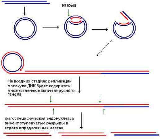
Fig. 11.4. Schematic of phage λ replication. Phage T-7 replication
Phage T-7 DNA is a linear double-stranded molecule (~40 kbp) with direct terminal repeats. Initiation of DNA Synthesis occurs within the molecule and proceeds bidirectionally. This yields two DNA molecules with incomplete 5’ ends. The 3’ ends of these molecules are complementary, allowing Complementary interaction between them (Fig. 5), after which the DNA strands can be covalently joined. At the next stage, a staggered cut is introduced, generating two DNA molecules with incomplete 3’ ends that are subsequently filled in (Fig. 11.5). Thus, full-length genomic DNA molecules are formed.
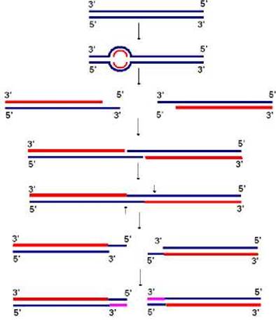
Fig. 11.5. Schematic of phage T-7 replication
Hepatitis B Virus Genome Replication
The hepatitis B virus DNA consists of a full-length (-) DNA strand (approximately 3.2 kb) with a protein attached to its 5’ end, and a shorter complementary (+) strand (1.7–2.8 kb). The (+) strand is complementary to both ends of the (-) strand and thus closes the (-) strand into a circle (Fig. 11.6).
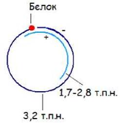
Fig. 11.6. Structure of hepatitis B virus DNA
The virion contains a virus-specific DNA polymerase capable of extending the (+) DNA. With its Participation in the infected cell, the viral DNA converts into a covalently closed circular form (Fig. 11.7).
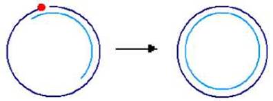
Fig. 11.7. Formation of double-stranded covalently closed DNA
RNA polymerase II, using the circular double-stranded DNA AS A template, synthesizes Two Types of +RNA:
a) short subgenomic +RNAs that serve as templates for Protein Synthesis;
b) long +RNAs encompassing the entire viral genome.
Long +RNAs serve as templates for DNA synthesis via virus-specific Reverse Transcriptase. First, the (-) DNA strand is synthesized. The reverse transcriptase uses a protein as a primer. Then, the same enzyme uses the (-) DNA strand to synthesize the (+) DNA strand.
Replication of phage φX174
The Genome of phage φX174 is represented by a single-stranded, covalently closed circular (+)DNA molecule. Upon entering the cell, it is converted into a circular, double-stranded, covalently closed DNA molecule with the participation of cellular primase, DNA polymerases I and III, and ligase. Subsequently, a phage-specific protein introduces a nick at a specific site in the (+)DNA strand and covalently attaches to the 5' end generated by the nick. The 3' end of the (+) strand resulting from the nick is used by cellular DNA polymerase as a primer, while the 5' end is displaced from the duplex, and the exposed (-)DNA strand acts as a template. After the Replication fork completes slightly more than one revolution, the phage-specific protein nicks the (+) strand at the genome boundary and attaches to the 5' end of the (+)DNA strand within the duplex. The displaced (+)DNA strand is circularized (Fig. 11.8). The released double-stranded DNA is again recruited into the synthesis of genomic (+)DNA. At the same time, the newly formed circular +DNA can be converted into double-stranded DNA and participate in the synthesis of new (+)DNAs or become incorporated into the virion.

Fig. 11.8. Schematic representation of phage φX174 replication
Replication scheme of Parvoviruses
The parvovirus genome is represented by single-stranded linear DNA. Both ends of the DNA strand are capable of forming hairpins due to the presence of self-complementary sequences.
Let us consider the replication of the adeno-associated virus (its reproduction is possible in the presence of an adenovirus).
Upon entry into the cell, the 3' end of the viral DNA forms a hairpin, which acts as a primer (Fig. 11.9). Then, synthesis of the complementary DNA strand takes place. As a result of this synthesis, reproduction of the complete viral genome is not achieved. Next, a virus-specific protein makes a nick in the parental strand. The resulting 3' end is used as a primer for the synthesis of the missing genome fragment. Consequently, a double-stranded DNA molecule is formed. Further, hairpins ("rabbit ears") are formed at both strands of one end of the DNA molecule, a primer is generated, and synthesis of the complementary strand begins. In this process, the DNA strand identical to the genome is displaced. The resulting double-stranded DNA can be further used for the synthesis of genomic single-stranded DNA via a similar mechanism (Fig. 11.9).

Fig. 11.9. Schematic representation of adeno-associated virus genome replication
Last update: 12/08/2026
Editorial and Educational Adaptation: This material has been compiled based on the primary/original source text. The project team performed an editorial review, corrected technical inaccuracies, structured sections, and adapted the content for an educational format.
What was processed:
- elimination of formatting defects (OCR errors, structural breaks, corrupted characters);
- editorial organization of content;
- standardization of terminology in accordance with academic sources;
- verification of factual statements against the original source text.
All mentions of the author, publication year, and origin of the primary text have been preserved in accordance with the source.