Anatomy of the Vessels and Nerves of the Trunk (Angioneurology) - Chornokulskyi S. T. 2011
Cardiovascular System
Vascularization of the Heart
The Main sources of Blood supply to The Heart are the right and left coronary Arteries (aa. coronariae dextra et sinistra). Secondary sources of blood supply include:
— pericardial and mediastinal Branches of the Thoracic Aorta (rr. pericardiaci et mediastinales);
— pericardiophrenic arteries (aa. pericardiacophrenicae), which are branches of the internal thoracic artery;
— smallest cardiac Veins (vv. cardiacae minimae), also known as the Thebesian Vascular System.
Class="center">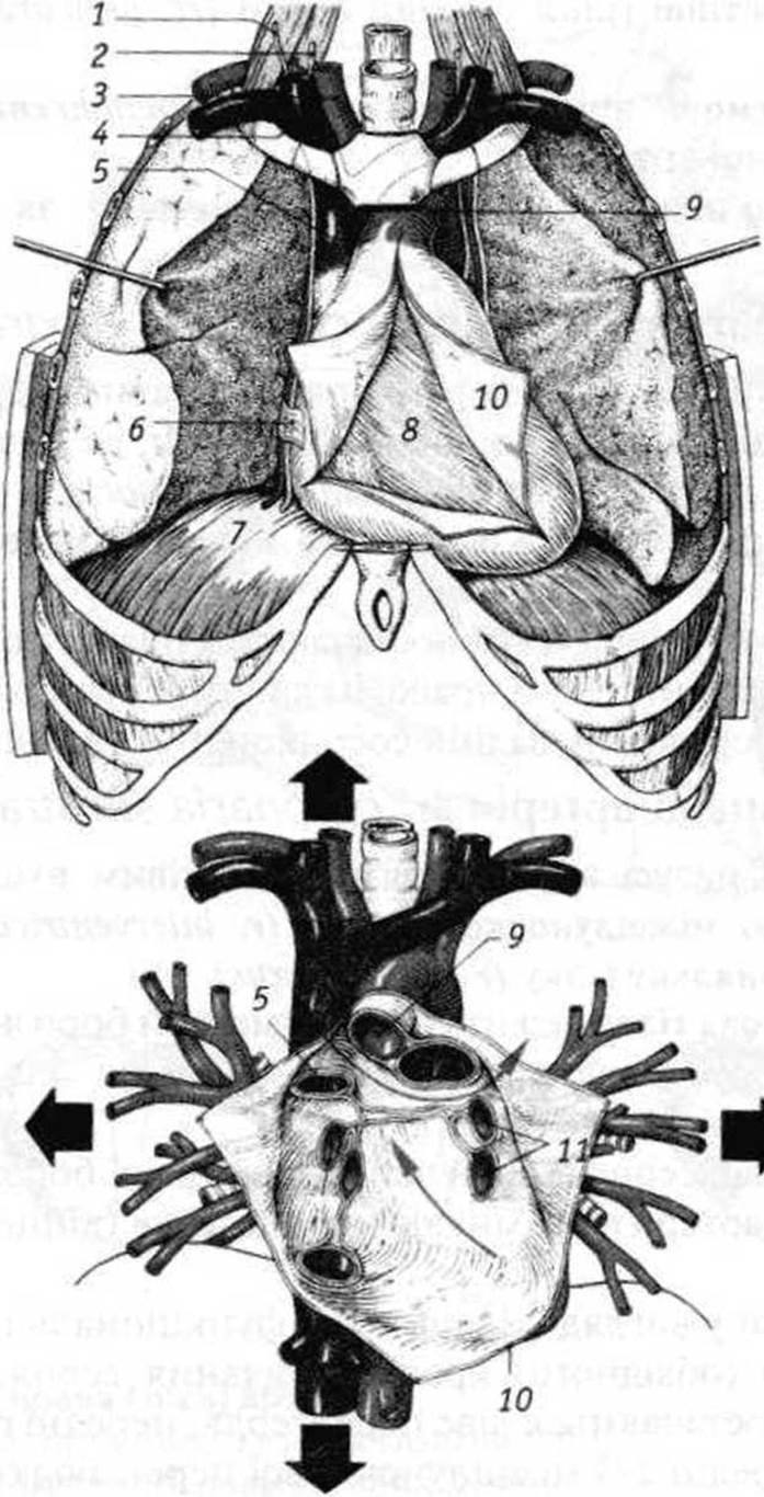
Fig. 18. Opened Pericardium (pericardial sac) and its topography:
1 - phrenic nerve (n. phrenicus); 2 - Vagus nerve (n. vagus);
3 - brachiocephalic trunk (truncus brachiocephalicus);
4 - right brachiocephalic vein (v. brachiocephalica dextra);
5 - SUPERIOR VENA CAVA (v. cava superior);
6 - mediastinal Pleura (pleura mediastinalis);
7 - Diaphragm (diaphragma);
8 - visceral layer (epicardium) [lamina visceralis (epicardium)];
9 - aorta (aorta);
10 - parietal layer of the pericardium (lamina parietalis pericardii);
11 - left Pulmonary veins (vv. pulmonales sinistrae)
Right coronary artery (a. coronaria dextra)
originates in the right aortic sinus, runs along the right segment of the coronary sulcus, and reaches the posterior interventricular sulcus, where it is referred to as the posterior interventricular branch (r. interventricularis posterior). Near the incisura of the cardiac apex, it anastomoses with the anterior interventricular branch (from coronaria sinistra).
The branches of the right coronary artery supply blood to the right atrium, the interatrial septum, the posterior walls of the right and left ventricles, the posterior third of the interventricular septum, and the posterior papillary Muscle of the left ventricle.
Left coronary artery (a. coronaria sinistra)
begins in the left aortic sinus, passes beneath the left auricle of the heart, where it divides into the anterior interventricular branch (r. interventricularis anterior) and the larger circumflex branch (r. circumflexus).
The anterior interventricular branch lies in the sulcus of the same name and, as noted above, anastomoses with the posterior interventricular branch, forming a longitudinal arterial hemiring (Fig. 19).
The circumflex branch courses around the heart within the left part of the coronary sulcus, anastomoses with the right coronary artery, and completes the transverse (coronary) arterial ring (Fig. 19).
These ring-shaped anastomoses serve as a functional adaptation for collateral blood supply to the heart. The branches of the left coronary artery supply the left atrium, the anterior walls of the left and right ventricles, the anterior 2/3 of the interventricular septum, and the anterior papillary muscle of the left ventricle.*
Various anatomical variations in the blood supply to the heart are also observed (Fig. 19, c), among which three main types are distinguished:
— right-coronary dominant
— left-coronary dominant
— balanced (intermediate).
The mid-right and mid-left types of heart blood supply are considered combinations of the main types with a significant predominance of the mid-right type.
* The names of the intraorgan Arteries of the heart correspond to the names of the regions, chambers, and structures of the heart: rr. atriales, rr. septales, rr. nodi, etc. Some of the myocardial branches are called "closing," meaning they are capable of blocking the blood supply to a specific area of the heart.
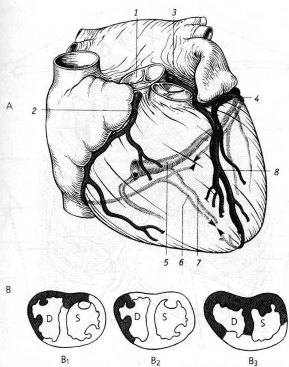
Fig. 19. Coronary (right and left) arteries:
A: 1 - posterior semilunar cusp of the aortic valve (valvula semilunaris posterior valvae aortae);
2 - right coronary artery (a. coronaria dextra);
3 - left coronary artery (a. coronaria sinistra);
4 - circumflex branch (r. circumflexus);
5 - coronary sinus (sinus coronarius);
6 - posterior interventricular branch (r. interventricularis posterior);
7 - middle cardiac vein (v. cordis media);
8 - anterior interventricular branch (r. interventricularis anterior);
B - types of heart blood supply:
B1 - balanced (intermediate); B2 - left-coronary dominant; B3 - right-coronary dominant
✵ dark field - right coronary artery;
✵ light field - left coronary artery
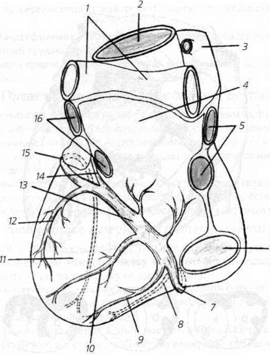
Fig. 20. VEINS OF THE Heart, posterior view (sketch the veins in blue on your own):
1 - pulmonary trunk (truncus pulmonalis) and left pulmonary artery (a. pulmonalis sinistra)
2 - ascending aorta (aorta ascendens);
3 - superior vena cava (v. cava superior);
4 - left atrium (atrium sinistrum);
5, 16 - pulmonary veins (right/left) (vv. pulmonales dextrae/sinistrae);
6 - INFERIOR VENA CAVA (v. cava inferior);
7 - small cardiac vein (v. cordis parva);
8 - anterior cardiac vein (v. cordis anterior);
9 - vein of the right ventricle (v. ventriculi dextri);
10 - middle cardiac vein (v. cordis media);
11 - left ventricle (ventriculus sinister);
12 - posterior vein of the left ventricle (v. posterior ventriculi sinistri);
13 - coronary sinus (sinus coronarius);
14 - oblique vein of the left atrium (v. obliqua atrii sinistri);
15 - great cardiac vein (v. cordis magna)
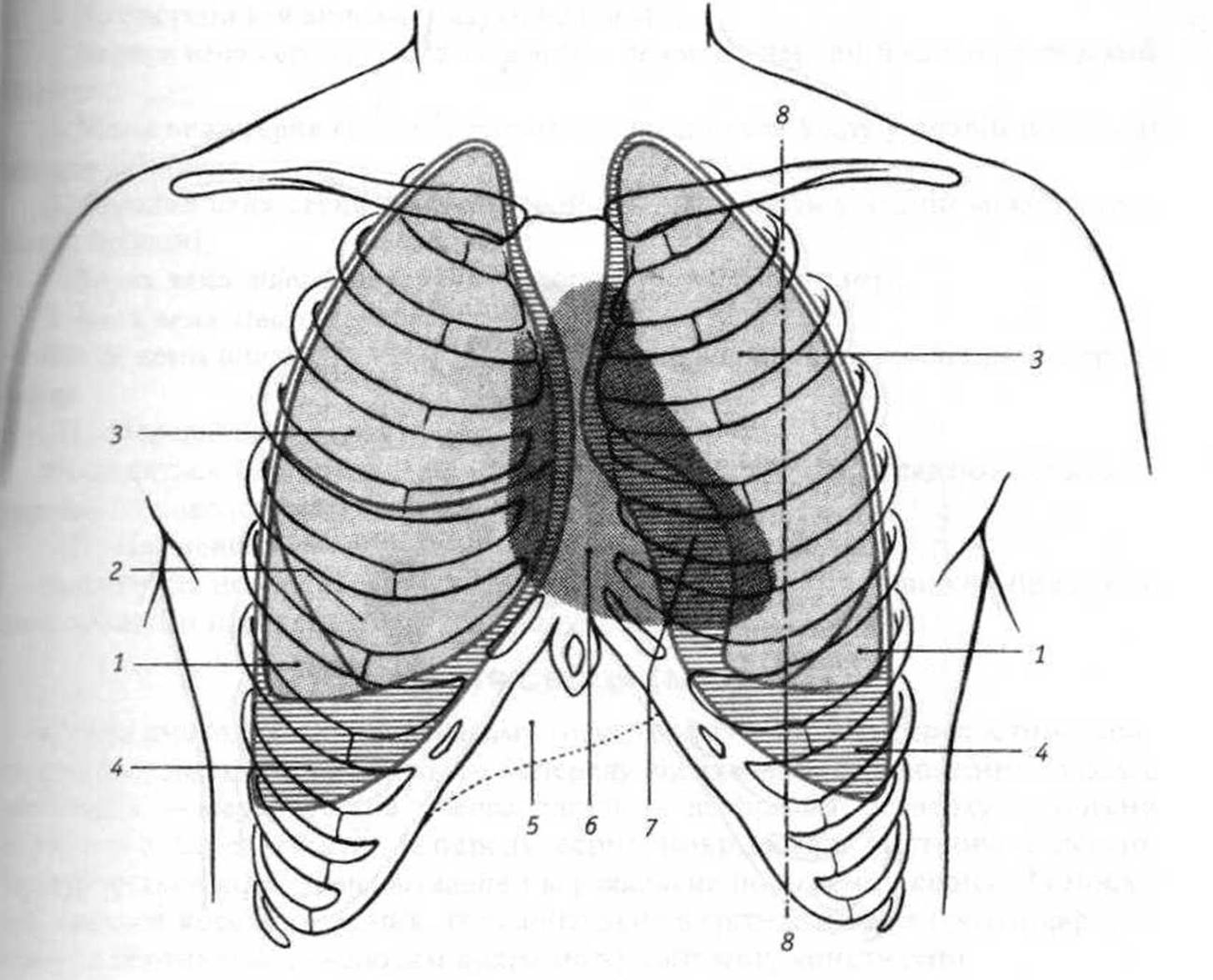
Fig. 21. Topography of the Heart, PROJECTION OF THE borders of the heart, Lungs, and pleura onto the anterior thoracic wall:
1 - inferior lobes of the lungs (lobі inferiores pulmones);
2 - middle lobe of the right lung (lobus medius pulmonis dextri);
3 - superior lobes of the lungs (lobi superiores pulmones);
4 - costodiaphragmatic recess (recessus costodiaphragmaticus);
5 - Liver (hepar);
6 - area of the heart not covered by the lungs;
7 - area of the heart covered by the lungs;
8 - left midclavicular line (linea medioclavicularis sinistra)

Fig. 22. Anatomical (see text) and auscultatory projection of the heart Valves:
1 - auscultatory projection of the mitral valve (apex of the heart);
2 - auscultatory projection of the tricuspid valve;
3 - auscultatory projection of the aortic valve;
4 - auscultatory projection of the pulmonary trunk valve.
Last update: 11/08/2026
Editorial and Educational Adaptation: This material has been compiled based on the primary/original source text. The project team performed an editorial review, corrected technical inaccuracies, structured sections, and adapted the content for an educational format.
What was processed:
- elimination of formatting defects (OCR errors, structural breaks, corrupted characters);
- editorial organization of content;
- standardization of terminology in accordance with academic sources;
- verification of factual statements against the original source text.
All mentions of the author, publication year, and origin of the primary text have been preserved in accordance with the source.