Pediatric Medical Genetics - S.I. Smiian 2003
Congenital malformations and hereditary diseases of the bronchopulmonary system in children
Most common malformations of the tracheobronchial system
Tracheobronchomegaly (Mounier-Kuhn syndrome, 1932) (Fig. 19) is a congenital enlargement of the Trachea and major Bronchi. In children, this condition is usually asymptomatic and is diagnosed via bronchoscopy and bronchography performed during secondary infections. Most commonly, however, tracheobronchomegaly presents between the ages of 30 and 50. Isolated forms are rare, occurring at an estimated rate of 1:100,000 (Minkov I.P., 1991). Clinically, it manifests as recurrent tracheobronchitis characterized by a bitonal irritant cough and prolonged sputum production. Treatment is similar to that of conventional endobronchitis. The prognosis for life is favorable.
Congenital lobar emphysema (synonyms: congenital localized emphysema, giant emphysema, tension emphysema) is characterized by the overinflation of a pulmonary lobe or segment and typically manifests in early infancy. It is a rare condition, with approximately 300 cases described by 1978. The pathology is associated with bronchial narrowing, aplasia, Dysplasia, or mucosal hypertrophy forming valve-like folds. Consequently, the volume of air entering the Lungs during inspiration exceeds that expelled during expiration (ball-valve mechanism).
Infants experience the most severe clinical distress in the first days of life, presenting with dyspnea, cyanosis, seizures, and loss of consciousness. Percussion reveals a hyperresonant (tympanic) note, while Auscultation shows diminished or absent breath sounds. Radiographic examination demonstrates a mediastinal shift toward the contralateral side.
The prognosis is unfavorable, and early mortality is high. In milder cases, the clinical course may be subacute or chronic. Affected children exhibit delayed physical growth and complain of a persistent cough and shortness of breath. It is important to note that mild forms of emphysema may be incidental findings. Chest radiography reveals increased radiolucency of the lung field, along with a depleted or absent pulmonary vascular pattern. Additionally, the Diaphragm appears flattened with restricted excursion, and a mediastinal hernia may be present.
Class="center">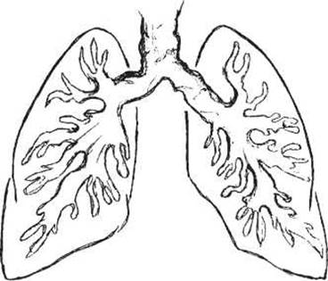
Fig. 19. Diagram of pathological Changes in the Trachea and Bronchi in Mounier-Kuhn syndrome.
Differential Diagnosis includes pneumothorax, pulmonary cysts, diaphragmatic hernias, foreign body aspiration-induced emphysema, and hypoplasia.
Treatment. Surgical intervention is indicated for tension syndrome resulting from mediastinal compression. Mild cases require conservative management, in which case lung GROWTH AND DEVELOPMENT are only minimally impaired.
Williams-Campbell syndrome (Fig. 20) is characterized by generalized Bronchiectasis caused by a structural defect in the bronchial Cartilage. First described in 1960, this morphological defect typically affects the segmental and subsegmental bronchi, predominantly in the lower lobes. As in lobar emphysema, the lung parenchyma remains well-aerated, leading to the hypothesis that these two conditions stem from a common underlying process. The prevalence is estimated at less than 1:100,000.
The Clinical presentation is dominated by bronchial obstruction and bronchopulmonary infection. Acute Pneumonia frequently develops During the first year of life, subsequently progressing to a chronic bronchopulmonary condition. Key symptoms include a persistent cough, dyspnea, hyperresonance on percussion, and dry or varying-caliber moist rales on auscultation. Other Clinical Features encompass a Pigeon chest (pectus carinatum), clubbing of the fingers, watch-Glass Nails, and impaired pulmonary ventilation. Radiography demonstrates an enhanced pulmonary pattern and signs of emphysema. Bronchoscopy reveals generalized bronchiectasis with ballooning dilation during inspiration and collapse with mucosal apposition during expiration.
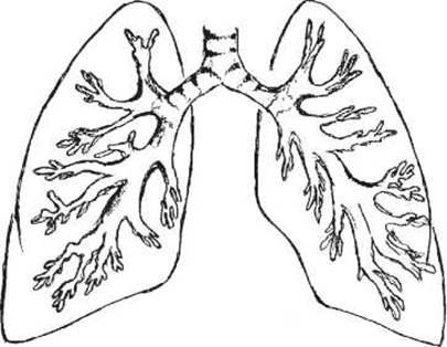
Fig. 20. Diagram of pathological changes in the trachea and bronchi in Williams-Campbell syndrome.
The prognosis is poor; the progressive course of the syndrome leads to Cor Pulmonale and cardiopulmonary failure, which is ultimately fatal.
Treatment is conservative.
Tracheoesophageal and bronchoesophageal fistulas manifest during the infant's first feeding with severe episodes of choking, coughing, and cyanosis. Uncorrected fistulas rapidly lead to severe, often fatal, aspiration pneumonia. This congenital anomaly is frequently associated with esophageal atresia.
Surgical treatment should be performed as early as possible. Prompt intervention offers a favorable prognosis for both health and life.
Last update: 11/08/2026
Editorial and Educational Adaptation: This material has been compiled based on the primary/original source text. The project team performed an editorial review, corrected technical inaccuracies, structured sections, and adapted the content for an educational format.
What was processed:
- elimination of formatting defects (OCR errors, structural breaks, corrupted characters);
- editorial organization of content;
- standardization of terminology in accordance with academic sources;
- verification of factual statements against the original source text.
All mentions of the author, publication year, and origin of the primary text have been preserved in accordance with the source.