Diagnosis and treatment of patients with recurrent gastroduodenal bleeding - Shaprynskyi V.O. 2009
Approaches to endoscopic diagnosis of the bleeding source and the capabilities of endoscopic hemostasis
In the hands of an experienced clinician, endoscopy is a highly informative diagnostic Procedure that can identify the source of upper gastrointestinal bleeding.
Preparation for endoscopic intervention and General Principles of its performance.
Active correction of hemodynamic parameters of Blood loss and associated syndromic disorders is the primary goal of intensive care in patients with acute gastrointestinal bleeding. It begins in the emergency department and continues, depending on the severity of the patient's condition, in the surgical or intensive care unit, on the endoscopy table, and in the operating room. Intensive care is an essential and integral part of preparing patients for endoscopy, as well as providing anesthetic support during the procedure.
Immediate preparation for upper gastrointestinal endoscopy consists of clearing the lumen and washing blood and clots from the mucosa of the Esophagus, Stomach, and duodenum. We believe that in most cases, this can be achieved by gastric lavage with ice-cold Water through a wide-bore nasogastric tube. The inner diameter of the tube allows for the evacuation of large clots, while local hypothermia helps reduce the intensity of bleeding or stop it completely. In recent years, however, we have advised against this procedure due to the high probability of washing away clots and restarting the Hemorrhage.
In most cases, endoscopy can be performed without any anesthetic support. However, anesthetics must be used in certain situations. A significant portion of these Procedures can be performed under local pharyngeal anesthesia with procaine, using opioid analgesics (1 ml of 2% promedol solution) and anticholinergics (1 ml of 0.1% atropine solution) for premedication. If patient agitation interferes with adequate visualization or hemostasis, intravenous sedation should be used more widely, or intravenous general anesthesia (endotracheal intubation in hemodynamically unstable patients). In cases of active gastric and duodenal peristalsis, the intravenous administration of antispasmodics (buscopan, papaverine, metacin, hexamethonium) is justified to induce relaxation.
Endoscopy has the following objectives:
1. Establishing an accurate Diagnosis (a. localization of the defect; b. status of hemostasis (e.g., ongoing bleeding, oozing from beneath a clot, or spurting hemorrhage); c. information regarding the source of bleeding (e.g., ulcer, tumor, erosion, etc.).
The stage of hemostasis is classified According to the universally recognized Forrest Classification:
1. Active bleeding (Forrest I)
- spurting (F Ia)
- oozing (F Ib)
2. Recent bleeding (Forrest II)
- non-bleeding visible vessel in the ulcer base (F IIa)
- adherent clot in the ulcer base (F IIb)
- flat pigmented spot (F IIc)
3. No signs of active bleeding, clean-based ulcer (F III).
The Institute of General and Urgent Surgery of the Academy of Medical Sciences of Ukraine has developed and utilizes a classification of hemostasis staging by V.T. Zaitsev et al. (1993):
Stage I. The stage of clot formation in the ulcer crater is characterized by arrested bleeding (often achieved through the aforementioned interventions for active hemorrhage) and a high risk of rebleeding.
Stage II. The stage of thrombus formation in the ulcer crater is characterized by arrested bleeding, ongoing thrombus consolidation, and a higher degree of hemostatic reliability compared to the previous stage. The thrombus in the ulcer crater is typically fixed and securely plugs both the ulcer crater and the vessels within it. Transition to the next stage occurs under METABOLISM/18.html">The Influence of gastric juice and local fibrinolytic factors, which can dissolve the thrombus, thereby reducing the reliability of hemostasis.
Stage III. The stage of thrombosed vessels in the ulcer crater is defined by the continuation of thrombolysis, which explains the more frequent occurrence of rebleeding at this stage, as well as the initial infiltration of the ulcer zone Tissues with plasma components.
Stage IV. The stage of fibrin formation in the ulcer crater is characterized by varying amounts of fibrin deposits within the crater, a lack of clear Definition of the ulcer crater itself, and a relatively high level of hemostatic reliability.
2. Providing prognostic information: the presence of signs of unstable hemostasis and ongoing bleeding (stigmata of recent hemorrhage (SRH)). SRH are most commonly found within the first 12-18 hours after the patient's admission. These signs of hemostasis are crucial for predicting rebleeding. A clean-based, fibrin-covered ulcer indicates a very low probability of recurrent bleeding; therefore, such patients are classified as low-risk and do not require prolonged hospitalization.
Early endoscopy, performed within the first 24 hours of admission, allows for a safe and rapid risk stratification of patients into high- and low-risk groups. Prompt identification of high-risk patients improves the quality of their management, while identifying low-risk patients helps reduce healthcare costs.
Therapeutic endoscopy
When choosing a specific method of endoscopic hemostasis, it is necessary, on the one hand, to consider its clinical efficacy in terms of achieving hemostasis and reliably preventing rebleeding, and on the other hand, to evaluate the method based on its technical simplicity, safety, availability, and cost. Given these characteristics and current clinical experience, we recommend having in the arsenal and utilizing for endoscopic hemostasis: monopolar and bipolar electrocoagulation, heater probe thermocoagulation, argon plasma coagulation; injection Methods using epinephrine, sclerosants, or tissue adhesives; and mechanical methods such as hemoclipping and band ligation. The Selection of a specific endoscopic hemostasis method or combination thereof for a given patient is primarily determined by the CHARACTERISTICS OF THE bleeding source and the Specific features of the method itself.
Class="center">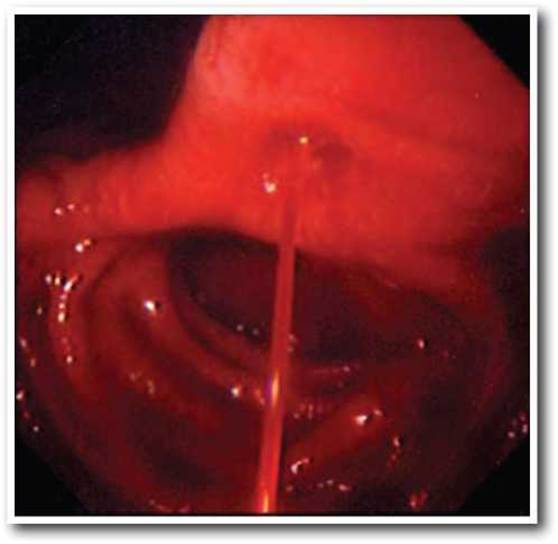
Fig. 9.1. Forrest Ia. Spurting bleeding.
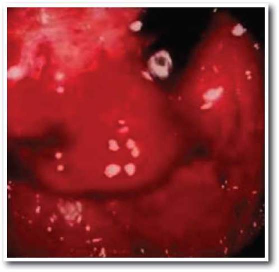
Fig. 9.2. Forrest Ib. Oozing of blood from the ulcer base (venous bleeding).
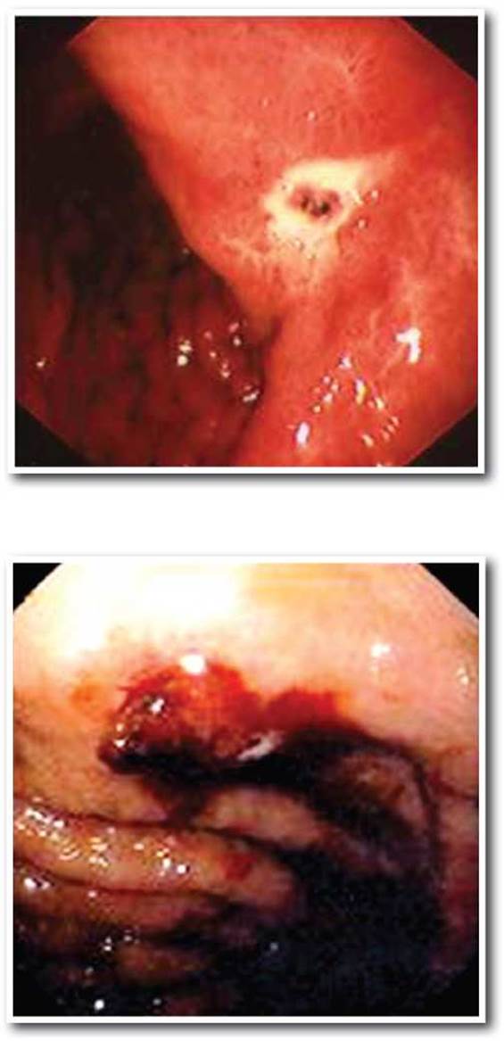
Fig. 9.3. Forrest IIa. Visible thrombosed vessel in the ulcer base.
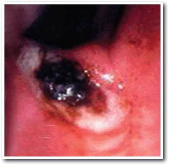
Fig. 9.5. Forrest IIb. Adherent clot in the ulcer base.
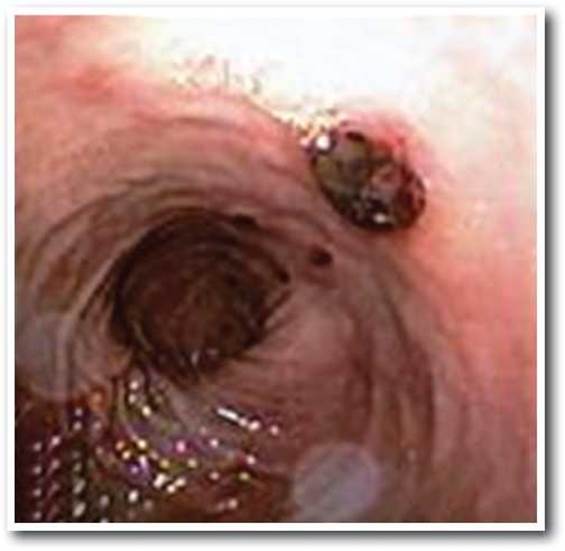
Fig. 9.6. Forrest IIc. Ulcer base covered with a flat pigmented spot.
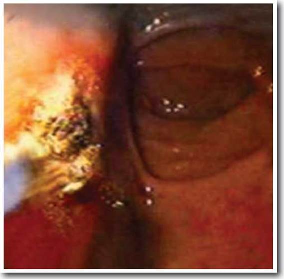
Fig. 9.8. Achieving endoscopic hemostasis using laser photocoagulation.
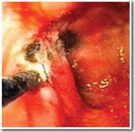
Fig. 9.9. Achieving endoscopic hemostasis using thermal cauterization.
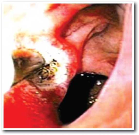
Fig. 9.10. Achieving endoscopic hemostasis using thermal cauterization.
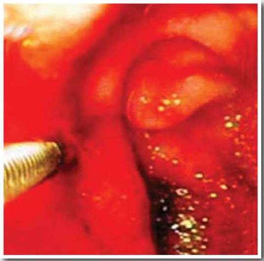
Fig. 9.11. Achieving endoscopic hemostasis using multipolar coagulation.
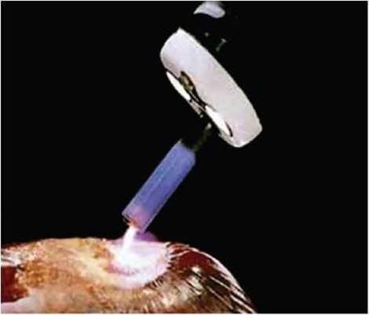
Fig. 9.12. Schematic illustration of argon plasma coagulation.
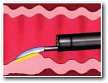
Fig. 9.13. Schematic illustration of argon plasma coagulation.
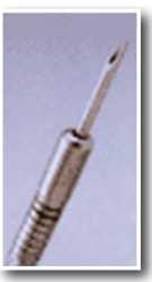
Fig. 9.14. Needle for endoscopic injection therapy.
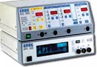
Fig. 9.15. Argon plasma coagulation device.
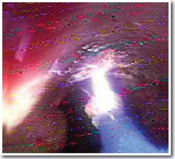
Fig. 9.16. Achieving endoscopic hemostasis using argon plasma coagulation.
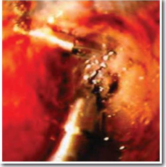
Fig. 9.17(a). Achieving endoscopic hemostasis using mechanical methods. Clipping of a vessel in the ulcer base.
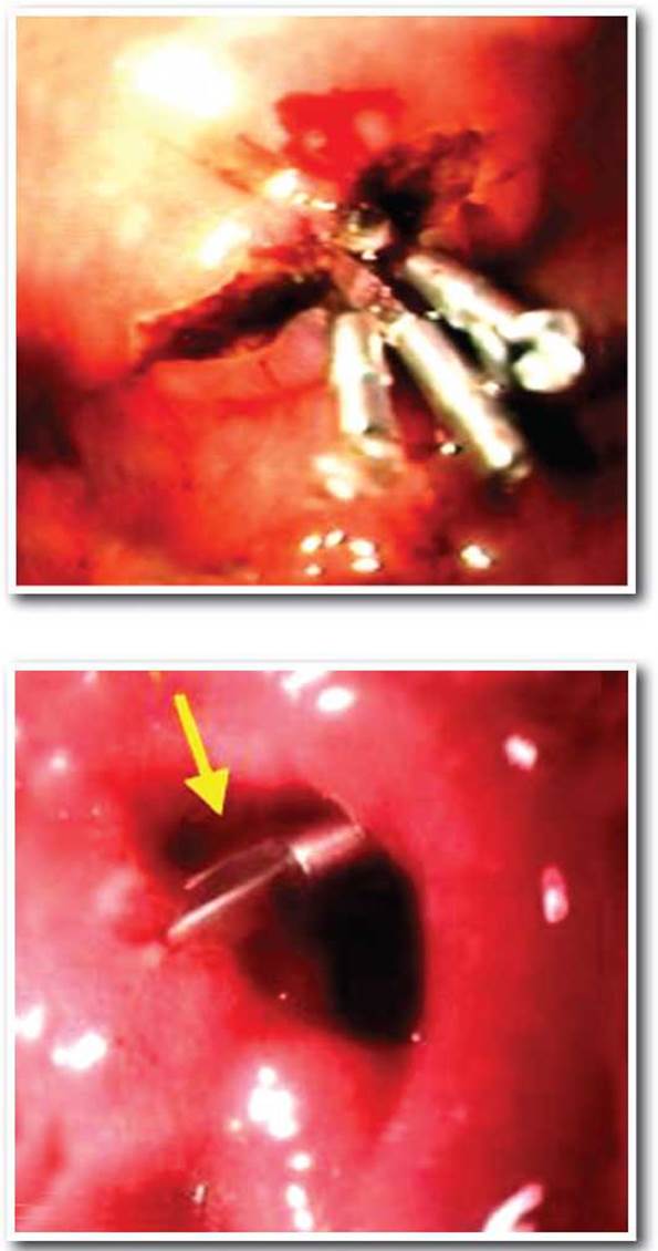
Fig. 9.17(b,c). Achieving endoscopic hemostasis using mechanical methods. Clipping of a vessel in the ulcer base
Numerous types of EHT are used, each aimed at arresting bleeding from a vessel. Therefore, it is crucial during endoscopic examination, prior to EHT, to irrigate the ulcer as thoroughly as possible to clear away debris and overlying clots. Naturally, this increases the likelihood of initiating active arterial bleeding; however, in such cases, it is quickly controlled using EHT.
Injection methods of endoscopic hemostatic therapy (EHT). These methods are extremely simple to use, widely applied, and currently the most cost-effective available. The Mechanism of hemostatic action lies in the vasoconstrictive effect of epinephrine, the tamponade effect (compression of the bleeding source by surrounding tissues), The Development of terminal arteritis, and a direct influence on the thrombus formation process at the site of the arterial defect.
The Role of tamponade in EHT was demonstrated by the injection of isotonic saline, where primary hemostasis was achieved in 80% of patients, with a recurrence rate of 29% in patients with ongoing bleeding. Epinephrine diluted 1:10,000 is frequently used for injection EHT. Typically, injecting 5-20 ml of epinephrine solution into four quadrants around the bleeding source is sufficient to achieve hemostasis. This controls active bleeding in over 90% of cases, though a high risk of recurrent bleeding (15-20%) is observed.
The Use of sclerosants or alcohol as monotherapy, or in combination with epinephrine, does not reduce The rate of recurrent bleeding, while introducing a risk of tissue necrosis at the injection site. Consequently, these techniques are currently rarely used. Other effective Materials for injection EHT include fibrin glue (a mixture of fibrinogen and Thrombin) and thrombin, which are highly effective and associated with a low complication rate. However, it has not been statistically proven that the use of fibrin glue, compared to epinephrine or their combination, is more effective. The difference lies solely in cost: 10 ml of epinephrine (1:10,000) costs £2.36, whereas 0.5 ml of fibrin glue costs £7.49 in the UK.
Thermal methods of hemostasis. These have the advantage of being deliverable directly to the bleeding source, providing a good coagulation effect, and being relatively inexpensive to use.
Electrocoagulation. Hemostasis is achieved through thermal action, i.e., coagulation of the bleeding source, resulting in its tamponade. EHT. Multipolar thermocoagulation (BiCAP) is widely used. Hemostasis is achieved due to eight separately positioned electrodes on the device surface.
Diathermocoagulation carries a certain risk of hollow organ perforation. The likelihood of this serious complication depends on The Nature of the bleeding source, the method of diathermocoagulation, current power, duration of exposure, and the intervention technique. In monopolar diathermocoagulation, one electrode (passive), in the form of a wide plate, is applied to the patient's external body surface (usually the thigh), while the other (active) is delivered through the endoscope's working channel to the target area. In bipolar and multipolar methods, all electrodes are located at the distal end of the probe, confining the action primarily to the tissue between them, without current spreading deeper into the substrate or through the patient's body. An electrode capable of delivering a conductive fluid enables hydrodiathermocoagulation, preventing coagulated blood from sticking to it and, consequently, avoiding recurrent bleeding when the probe is withdrawn from the bleeding source. The targeted fluid jet delivery function through the electrode is absolutely indispensable when the procedure is performed using an endoscope without a dedicated irrigation channel.
Thermocautery (cauterization or thermal coagulation) (heater probe (HP)) is currently considered the method of choice for endoscopic hemostasis. Unlike diathermocoagulation, the active element of thermocautery is not electric current itself, but the working tip of the thermal probe heated by it to over 100 C. This 'mini-soldering iron' is used to cauterize the vessel, resulting in a hemostatic effect. Because the thermal probe tip is Teflon-coated, it is less prone to sticking to the blood clot, while retaining all the advantages of the contact method. A pressurized fluid jet simultaneously cools the probe and washes away accumulated blood from the bleeding source, creating more favorable conditions for cauterization. To stop ongoing bleeding, it is advisable to press the probe tip directly against the bleeding vessel, temporarily halting the hemorrhage, and then initiate heating of the thermal probe's working HEAD. To arrest ongoing bleeding, a substantial amount of energy is applied (20-30 Joules), whereas lower energy parameters (10-20 Joules) are used to coagulate thrombosed vessels and create a more durable coagulum.
The heating level of the thermal probe is easily metered and regulated; when basic safety rules are followed, it does not cause deep Burns and is successfully used in virtually all types of non-variceal gastrointestinal bleeding.
Multipolar coagulation and heater probe (HP) require the continuous availability of power generators. The energy supplied by the generators passes through a conductor and is converted into thermal energy directly at the bleeding source. The maximum Temperature for multipolar coagulation is 100o C and for HP is 250 oC. In comparative studies, neither method has shown superiority over the other. However, thermocautery is most commonly used abroad. The typical voltage applied to the tip is 20 volts, and the risk of perforation is low.
Lasers, such as the neodymium laser, are also used for thermocoagulation. However, using a neodymium laser is challenging during EHT, making the experience of the endoscopist crucial, and the presence of an assistant is required. The cost of such equipment is high, so its use is only practical in large medical centers.
Argon plasma coagulation is based on coagulation using a stream of argon gas. The first encouraging results obtained in endoscopic hemostasis and Prevention of recurrent bleeding using a jet of ionized argon ('argon plasma') place this method among the most effective and clinically proven means of hemostasis. One of the key advantages of this method is that it is non-contact, thereby avoiding the side effects inherent in contact techniques, such as recurrent bleeding due to clot avulsion. Argon plasma can be used for hemostasis in hard-to-reach areas (for example, in a deformed duodenal bulb) due to its affinity for high-conductivity zones (liquid blood and fresh clots). Importantly, the technique has virtually no tissue-ablating effect: argon plasma coagulation minimally vaporizes and damages tissue, and instead of 'burning out' the ulcer defect, it seals it by forming a dense protective layer. The coagulating effect of argon plasma is easily metered; it does not exert a pronounced thermal effect on the deep layers of the intestinal wall (the depth of argon plasma penetration into the tissue is no more than 2-3 mm) and is therefore safe regarding organ perforation.
To perform argon plasma coagulation, a special probe connected to an APC-300 argon source and an ICC-200 electrocoagulator is passed through the endoscope's biopsy channel. The distal end of the probe is positioned 10 mm from the endoscope tip and 5-10 mm from the substrate. At an argon flow rate of 2 L/min and an electrical power of 60 W, the substrate surface is coagulated with 4-5 pulses of several seconds each until reliable (endoscopically confirmed) hemostasis is achieved. If a fixed clot is present, the vessel base must be cleared by targeted irrigation, and hemostasis performed according to the described method.
Initial clinical experience has shown that the use of argon plasma is particularly effective in bleeding from vascular aneurysms, Dieulafoy's lesions, telangiectasias, etc., where it is necessary to provide a powerful hemostatic effect without excessive destruction of the mucous membrane. However, argon plasma coagulation is finding increasingly widespread use in bleeding of ulcer Etiology, tumors, and Mallory-Weiss syndrome.
However, this method is insufficient to arrest spurting bleeding. Using this method is hazardous when manipulating large-diameter non-bleeding vessels, as argon plasma coagulation provides only a superficial burn. Recent studies have shown that the use of epinephrine in combination with argon plasma coagulation achieves primary hemostasis in 98.1% of cases, though recurrent bleeding occurs in 10%.
Mechanical methods of hemostasis. Hemoclips can be applied to visible vessels, and although placement can sometimes be challenging, particularly for ulcers located in hard-to-reach areas, this method can be considered the best for EHT in patients with PEPTIC ULCER DISEASE complicated by acute gastrointestinal bleeding. It is well known that injection or thermal EHT is insufficiently effective for Arteries in the ulcer base wider than 1 mm, whereas a properly applied clip can arrest bleeding from relatively large-diameter vessels.
Nishiaki, Lin HY, Chung CH et al. conducted a large study comparing EHT monotherapy using epinephrine, thermocautery, and mechanical clipping. The study data showed that all three methods are equally effective in achieving primary hemostasis. However, the rate of recurrent bleeding is lower with the application of hemoclips. Achieving hemostasis using a neodymium laser or clip placement requires specific skills from the endoscopist, especially when ulcers are located in hard-to-reach areas.
Combination of different EHT methods. Combined use of hemostatic methods is quite common in daily clinical practice. The application of various EHT methods is well-founded, as different EHT modalities are based on distinct mechanisms of achieving hemostasis. Their most frequent combination during primary endoscopic intervention is the sequential use of the injection method (vasoconstrictors) and thermal methods. By complementing each other, they highlight their positive aspects while mitigating the drawbacks inherent in each method. Overall, this enables more effective, reliable, and durable hemostasis.
Causes of EHT failure. Along with the growing number of methods to achieve primary hemostasis and the increasing skills of endoscopists, repeat EHT in case of recurrent bleeding is undoubtedly indicated for all patients with GIB, as it can significantly improve patient outcomes. Complications after EHT (organ perforation, development of cicatricial strictures), even repeat procedures, occur in an extremely low percentage of cases and arise mainly after the injection of sclerosants and alcohol, or when monopolar thermocoagulation is used. Two large studies showed that EHT failure was observed predominantly in patients with active ongoing bleeding, giant ulcers (> 2cm), and ulcers located on the posterior wall of the duodenum.
The results of repeat EHT Treatment are contradictory. In his studies, Lau JYW showed primary hemostasis failure in 1.5% of cases, which were managed with emergency surgery. However, the study concluded that routine second-look endoscopy is not recommended for daily practice.
At present, it is well understood only that the approach to treating patients with GIB must be based on individual endoscopic findings, the patient's overall condition, and the presence of comorbidities.
We studied a group of 56 patients with gastrointestinal bleeding of peptic ulcer etiology. To achieve primary hemostasis, endoscopic clipping of the vessel at the ulcer base and infiltration of the vessel at the ulcer base were used. The indications for EHT were: I - ongoing bleeding, and II - a visible thrombosed vessel and the presence of a loose, fresh clot at the ulcer base.
Table 6.2.1. Treatment outcomes of patients with PUD complicated by GIB using endoscopic bleeding control techniques.
|
Group I |
Group II |
||||||
|
F 1b |
F 2a |
F 2b |
|||||
|
total |
recurrence |
total |
recurrence |
total |
recurrence |
||
|
EIT |
0.001% epinephrine soln. |
4 |
1 |
6 |
1 |
0 |
0 |
|
0.9% NaCl soln. |
1 |
0 |
2 |
0 |
2 |
0 |
|
|
Alcohol (70%) - novocaine (0.5%) mixture 1:1 |
3 |
2 |
6 |
1 |
11 |
1 |
|
|
Endoscopic clipping |
4 |
2 |
14 |
3 |
3 |
0 |
|
|
Total |
12 |
5 |
28 |
5 |
16 |
1 |
|
Among the methods of endoscopic hemostasis, we used clipping of the vessel at the ulcer base, as well as endoscopic injection therapy (EIT) with 0.001% epinephrine solution, 0.9% sodium chloride solution, and a 1:1 mixture of alcohol (70%) and novocaine (0.5%). In Group I, EHT was performed to stop bleeding, while in Group II, it was used to prevent bleeding recurrence. Recurrence occurred in 11 (19.6%) patients across both groups: in 5 (41.7%) cases among Group I patients, and in 6 (13.6%) cases in Group II. When studying the development of recurrent bleeding depending on the specific EHT method used, we see that in cases of ongoing bleeding, the best hemostatic effect was achieved in the subgroup that received endoscopic injection therapy with 1:10000 epinephrine (25% recurrence rate). In Group II patients, infiltration of the ulcer with an alcohol-novocaine mixture yielded the best results (11.7% recurrence rate).
Fatal outcomes were observed only in Group I patients (2 patients, 16.6%). Notably, both patients experienced recurrent bleeding, which caused the death of one patient, while the other died due to decompensation of comorbidities against the Background of severe post-hemorrhagic anemia.
The overall mortality rate among patients who underwent EHT was 3.6%. Among patients in both groups, Surgical treatment was performed in 2 patients from Group I. Ulcer excision followed by duodenoplasty was performed at the height of recurrent bleeding.
We present the following clinical case.
Patient K., 33 years old, Case History No. 8429, was admitted to the clinic of hospital surgery on an emergency basis on May 10, 2004. BP - 140/90 mmHg, Ps - 100 bpm. Hb - 62 g/l, erythrocytes - 2.2*109/l, CI - 0.9. According to the medical history, the onset of illness was on May 9, 2004, when vomiting of fresh blood occurred after a dietary indiscretion, followed by melena the next day. The patient was hospitalized in the abdominal surgery department of the hospital surgery clinic, where an EGD was performed. The duodenal bulb was hyperemic; on the anterior-inferior wall, there was a flat ulcer up to 1 cm in diameter, covered with fibrin, with a large thrombosed vessel in the center. The clot was fresh and loose. Endoscopic injection therapy with 1:10000 epinephrine solution was performed. The bleeding was controlled. The patient was diagnosed with: Peptic ulcer disease. Duodenal ulcer complicated by bleeding (F IIa).
Severe post-hemorrhagic anemia. The patient received conservative hemostatic, antiulcer, infusion, and blood Transfusion Therapy. However, on May 14, 2004, recurrent bleeding occurred. BP - 90/40 mmHg, pulse - 120 bpm, multiple episodes of melena. The patient was urgently taken to the operating room. During the surgical intervention, a duodenal bulb ulcer located on the anterior-inferior wall, up to 1 cm in diameter, was found. In the center of the ulcer crater, there was a vessel with ongoing spurting bleeding. The vessel was suture-ligated. Ulcer excision, duodenoplasty, and selective vagotomy were performed. The wound was closed in layers.
In the postoperative period, the patient received antibacterial drugs, antisecretory agents, infusion and blood transfusion therapy, and analgesics.
Laboratory parameters: (10.05.2004) Hb - 62 g/l, erythrocytes - 2.2*109/l, CI - 0.9, leukocytes - 6.5*109/l, total protein 52 g/l; total bilirubin 6.8 µmol/l; urea 5.0 mmol/l; K+ - 4.5 mEq/l, Na+ - 143 mEq/l, glucose - 5.1 mmol/l, fibrinogen - 3.9 g/l, PTI - 77%, ALT - 0.12 mmol/l.
Laboratory parameters: (14.05.2004) Hb - 50 g/l, erythrocytes - 1.41*109/l, CI - 1.06, leukocytes - 9.0*109/l, total protein 49.3 g/l; total bilirubin 18 µmol/l; urea 9.1 mmol/l, glucose - 8.0 mmol/l, fibrinogen - 3.55 g/l, PTI - 84%.
Laboratory parameters: (24.05.2004) Hb - 90 g/l, erythrocytes - 3.3*109/l, CI - 0.8, leukocytes - 11.0*109/l.
The postoperative period was uneventful, and the patient was discharged from the clinic in satisfactory condition on May 28, 2004.
Thus, summarizing all of the above, we can conclude that primary endoscopic hemostatic therapy is indicated for all patients presenting with signs of ongoing bleeding and unstable hemostasis upon admission for gastrointestinal bleeding of peptic ulcer etiology. However, recurrent bleeding after achieving hemostasis with endoscopic methods occurred in 41.7% of patients with diffuse oozing bleeding from the ulcer (F 1b). In patients with unstable hemostasis (F 2a - F 2b), recurrence occurred in 13.6% of cases. Therefore, attempts at endoscopic control of ongoing bleeding often prove ineffective and lead to delayed surgery; thus, they should be considered temporary measures during patient preparation for surgery. Nevertheless, endoscopic hemostatic therapy in peptic ulcer disease complicated by acute gastrointestinal bleeding can serve as an alternative to surgical intervention in patients at high risk of potential fatal outcomes.
Last update: 11/08/2026
Editorial and Educational Adaptation: This material has been compiled based on the primary/original source text. The project team performed an editorial review, corrected technical inaccuracies, structured sections, and adapted the content for an educational format.
What was processed:
- elimination of formatting defects (OCR errors, structural breaks, corrupted characters);
- editorial organization of content;
- standardization of terminology in accordance with academic sources;
- verification of factual statements against the original source text.
All mentions of the author, publication year, and origin of the primary text have been preserved in accordance with the source.