Review of Medical Physiology - William F. Ganong 2002
Physiology of Nerve and Muscle Cells
Synaptic and Junctional Transmission
Neuromuscular Transmission — Nerve Terminals in Smooth and Cardiac Muscle
Postganglionic fibers of Neurons innervating smooth Muscles branch extensively and maintain close contact with Muscle Cells (Fig. 4-36). Some of these nerve fibers contain clear vesicles and are cholinergic, whereas others contain characteristic electron-dense vesicles filled with norepinephrine. Neither end plates nor other specialized postsynaptic structures are found in this type of junction. The nerve fiber runs along the membranes of the muscle cells, occasionally indenting them. Numerous branchings of noradrenergic, and presumably also cholinergic, neurons form swellings (varicosities) that lack Schwann Cell covering and contain synaptic vesicles (see Fig. 4-36). A single noradrenergic neuron features over 20,000 varicosities spaced approximately 5 µm apart. Each varicosity presumably releases a neurotransmitter, meaning that transmission occurs at multiple sites along each axon. This arrangement enables a single neuron to innervate A large number of effector cells. A junctional type in which a neuron forms synapses On the surface of another neuron or a smooth muscle cell and continues onward to establish similar contacts with other cells is termed a synapse en passant.
Class="center">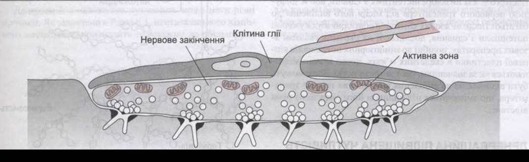
Fig. 4-35. Neuromuscular Junction. Note that clear vesicles containing acetylcholine cluster at the active Zones of the nerve terminal. These zones are located opposite the junctional folds of the motor end plate (reproduced with permission from Dowling JE: Neurons and Networks: An Introduction to Neuroscience. The Belknap Press of Harvard University Press, 1992).
In The Heart muscle, cholinergic and noradrenergic fibers terminate on the sinoatrial node, atrioventricular node, and bundle. Noradrenergic fibers also innervate the ventricular musculature. The exact structure of nerve endings on nodal tissue remains unclear. In the ventricles, contacts between noradrenergic fibers and cardiac muscle fibers are similar to those observed in smooth muscle.
Electrical Responses
In smooth muscles where noradrenergic input is excitatory, stimulation of noradrenergic nerves triggers intermittent partial depolarizations resembling miniature end-plate potentials, known as excitatory junction potentials (EJPs). Summation of these potentials occurs upon repetitive stimulation. Similar EJPs have been identified in Tissues responsive to excitatory cholinergic input. Conversely, in tissues where adrenergic input is inhibitory, stimulation of noradrenergic nerves generates inhibitory junction potentials (IJPs).
Electrical responses of this type have been observed in numerous Smooth Muscle Tissues following single-nerve stimulation, though their latencies vary. This Variability is presumably due to the synapse en passant arrangements described above, and may also be explained by the presence of low-resistance junctions between smooth muscle cells or the diffusion of the neurotransmitter from its release site to multiple smooth muscle cells. Miniature excitatory junction potentials recorded in isolated smooth muscle preparations resemble miniature end-plate potentials in Skeletal Muscle, yet they differ significantly in both amplitude and duration. They likely represent responses to the release of individual quanta of transmitter, modified by varying diffusion distances.
Last update: 10/08/2026
Editorial and Educational Adaptation: This material has been compiled based on the primary/original source text. The project team performed an editorial review, corrected technical inaccuracies, structured sections, and adapted the content for an educational format.
What was processed:
- elimination of formatting defects (OCR errors, structural breaks, corrupted characters);
- editorial organization of content;
- standardization of terminology in accordance with academic sources;
- verification of factual statements against the original source text.
All mentions of the author, publication year, and origin of the primary text have been preserved in accordance with the source.