Operative Surgery and Topographic Anatomy - 2016
Content Module No. 3. Topographic Anatomy and Operative Surgery of the Abdominal Wall and Abdominal Cavity Regions
Topic 11. Inguinal Region. Inguinal Canal, Operations for Inguinal Hernias. Surgical Treatment of Congenital, Strangulated, and Sliding Hernias
1. Relevance of the topic: To understand The Mechanism of inguinal hernia formation, it is necessary to study the anatomical, physiological, age, and sex-related Features of the inguinal region Structure, and to select an appropriate method for Inguinal Canal plasty.
2. Specific objectives:
1. Explain the Anatomy of the inguinal region: boundaries, external landmarks, and layered topography.
2. Analyze the anatomy of the inguinal triangle as a weak spot in the musculo-aponeurotic layer.
3. Explain the anatomy of the posterior surface of the anterior abdominal wall and the inguinal region (peritoneal fossae and folds).
4. Explain the Surgical anatomy of indirect and direct hernias.
5. Explain how to perform the most common Methods of herniorrhaphy and inguinal canal plasty.
3. Tasks for independent work during preparation for the Class.
3.1. List of key terms, parameters, and characteristics that students must master when preparing for the class:
Term |
Definition |
1. Bassini herniorrhaphy 2. Girard-Spasokukotsky herniorrhaphy 3. Martynov herniorrhaphy |
1. A surgical method for treating inguinal hernias involving the plasty of the posterior wall of the inguinal canal 2. A surgical method for treating inguinal hernias involving the plasty of the anterior wall of the inguinal canal 3. A surgical method for treating inguinal hernias by forming a duplication of the external oblique abdominal Muscle aponeurosis, wherein the upper flap of the aponeurosis is sutured to the inguinal ligament and the lower flap is placed over it (without suturing the Muscles) |
3.2. Theoretical questions for the class:
1. What are the inguinal triangle and the inguinal space?
2. Where and how are the superficial and deep inguinal rings projected?
3. What structures form the folds and fossae on the internal surface of the anterior abdominal wall?
4. How is the Spermatic Cord positioned relative to the hernia sac in Direct and Indirect inguinal hernias? What anatomical structures compose the spermatic cord?
5. How should the Skin incision be performed in direct and indirect inguinal hernias?
6. How is inguinal canal plasty performed according to Girard-Spasokukotsky, Kimbarovskiy, Martynov, and Bassini?
7. Is it possible to operate on an incipient hernia (in a child) without opening the inguinal canal?
3.3. Practical skills to be acquired during the class:
1. Identification of the BOUNDARIES OF THE inguinal region, external and internal landmarks, and projections: pubic tubercle, superficial inguinal ring, course of the inguinal ligament, and borders of the internal oblique and transversus abdominis muscles.
2. Management of the hernia sac and plasty of the inguinal canal walls using various methods: Girard-Spasokukotsky, Martynov, Bassini, and their modifications (Kimbarovskiy sutures).
4. Content of the topic
At the beginning of the class, the instructor verifies students' understanding of the topic and the practical tasks through an oral quiz and by monitoring their independent preparation.
Next, under the Supervision of the instructor, the student group begins hands-on work with the material: using anatomical tables, a skeletal model, and a cadaver, they outline the boundaries of the inguinal region and identify external landmarks: the anterior superior iliac spine, pubic tubercle, Pubic Symphysis, inguinal ligament, and the lateral margin of the rectus abdominis muscle. They then project the inguinal triangle, bounded inferiorly by the inguinal ligament, medially by the lateral edge of the rectus abdominis muscle, and superiorly by a horizontal line drawn through the point located at the junction of the middle and lateral thirds of the inguinal ligament. Within this triangle lie the inguinal canal along with its superficial and deep rings.
The students then proceed to dissect the walls of the inguinal canal and the spermatic cord. To do this, medial to the pubic tubercle, they locate the exit point of the spermatic cord—the superficial inguinal ring. From this point, the aponeurosis of the external oblique muscle of the abdomen (the anterior wall of the inguinal canal) is incised upward for 5-6 cm. The edges of the aponeurosis are retracted superiorly and inferiorly to expose the lower edge of the internal oblique and transversus abdominis muscles (the superior wall of the inguinal canal). The spermatic cord is isolated and retracted inferiorly. The inguinal ligament is exposed in the shape of a trough (the inferior wall of the inguinal canal). The tense fascial sheet extending between the inguinal ligament and the posterior surface of the transversus abdominis muscle—forming the posterior wall of the inguinal canal—is the transversalis fascia.
Attention is drawn to the fact that the area of the transversalis fascia not covered by muscles has a triangular or oval shape and is referred to as the inguinal triangle (Hasselbach's triangle). Students examine the correlation between the width of the inguinal space and the degree of muscular development (variations in the shape of the inguinal space). The site in the transversalis fascia where the spermatic cord enters the inguinal canal is called the deep inguinal ring, which typically corresponds to the lateral inguinal fossa.
From the peritoneal cavity side, the medial and lateral inguinal fossae are identified on the Peritoneum of the anterior abdominal wall. The students compare the projections of the peritoneal inguinal fossae with the superficial and deep inguinal rings.
The instructor highlights the topographical features of the ilioinguinal nerve, which lies on the superior wall of the inguinal canal and upon the elements of the spermatic cord. This anatomical detail must be taken into account during hernia repair. Students identify the key elements of surgical hernia anatomy: the hernia ring, the coverings of the hernia sac, and the hernia contents. In this context, the Classification and variety of inguinal hernias are discussed: direct, indirect, congenital, acquired, sliding, and incarcerated. Special emphasis is placed on the characteristics of direct and indirect inguinal hernias, highlighting differences in the Location OF THE hernia rings, the relationship to the hernia sac and elements of the spermatic cord, and the association with congenital developmental anomalies (congenital inguinal hernia) (Fig. 49).
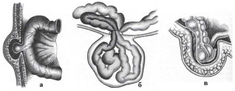
Fig. 49. Types of hernias.
a - partial (Richter's) incarceration of the intestine; b - retrograde incarceration; c - extraperitoneal sliding hernia of the cecum.
Subsequently, divided into surgical teams, the students perform a hernia repair Procedure. They justify the surgical approach and emphasize the necessity of incising the inguinal canal along its entire length. They focus on individual Stages of the surgical technique: management of the hernia sac and plastic repair of the hernia ring according to Girard-Spasokukotsky, Martynov, and Bassini. The contributions of prominent surgeons to herniology (Spasokukotsky, Bobrov, Krymov, Martynov, Kimbarovsky, Kukudjanov) are highlighted.
The possibility of performing herniorrhaphy without opening the inguinal canal for early-stage hernias in children is also discussed, referencing the Roux, Opel, and Krasnobayev methods.
SURGICAL Procedures FOR INGUINAL HERNIAS
Bassini Method
The patient is placed in a supine position; occasionally, the Trendelenburg position is used for large hernias. Local anesthesia is administered. An oblique incision 8–12 cm in length is made roughly parallel to the inguinal ligament over the site of maximum protrusion of the hernia sac, dividing the skin, subcutaneous tissue, and superficial fascia. Bleeding transected vessels (superficial epigastric and superficial external pudendal Arteries) are ligated. Using a grooved director inserted into the superficial opening of the inguinal canal, the aponeurosis of the external oblique abdominal muscle is incised. Its margins are retracted laterally, the hernia sac is excised, and the posterior wall of the inguinal canal is reconstructed. The spermatic cord is elevated on a gauze tape, and the inferior edge of the internal oblique and transversus abdominis muscles is sutured to the inguinal ligament.
Sutures placed at the upper angle of the wound must not compress the spermatic cord, and the opening around it should comfortably admit the tip of a little finger. The spermatic cord is placed back onto the newly formed muscular bed, over which the edges of the incised external oblique aponeurosis are sutured, followed by skin closure.
Girard-Spasokukotsky Method
Unlike the Bassini procedure, the Girard method focuses on the reconstruction of the anterior wall of the inguinal canal.
The lower edges of the internal oblique and transversus abdominis muscles are sutured to the inguinal ligament superior to the spermatic cord. Next, the upper edge of the external oblique aponeurosis is sutured to the inguinal ligament, after which the lower edge is overlapped on top of it (like the flaps of a coat) and secured with several interrupted sutures. As a result, a duplication (double-breasted fold) of the external oblique aponeurosis is formed in the region of the inguinal triangle. In the Spasokukotsky modification, the upper edge of the external oblique aponeurosis flap and the edges of the internal oblique and transversus abdominis muscles are sutured together simultaneously, and then the lower edge of the aponeurotic flap is laid over them as a second layer (duplication). The Martynov method creates a duplication of the external oblique aponeurosis by suturing the upper aponeurotic flap to the inguinal ligament and overlaying the lower flap on top of it (without suturing the underlying muscles) (Figs. 50, 51, 52, 53).
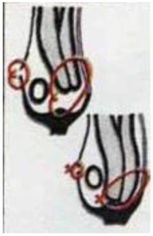
Fig. 50. Inguinal canal repair according to Bassini.
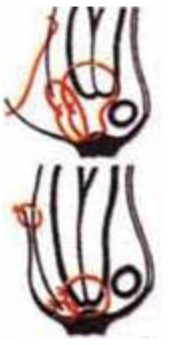
Fig. 51. Inguinal canal repair according to Girard.
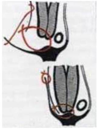
Fig. 52. Inguinal canal repair according to Girard-Spasokukotsky.

Fig. 53. Plastic repair of the inguinal canal according to Kimbarovsky.
5. Self-assessment Materials
A. Self-assessment tasks
Test No. 1
During surgery for an inguinal hernia, the surgeon retracted the Contents of the inguinal canal. What constitutes the contents of this canal in a male?
a. Inguinal ligament
b. Spermatic cord
c. Inferior epigastric artery
d. Round ligament of the Liver
e. Round ligament of the Uterus
Test No. 2
Which peritoneal fold is located above the inferior epigastric artery?
a. Median
b. Medial
c. Lateral
d. Superior
e. Inferior
Test No. 3
A patient has developed a hernia in the medial inguinal fossa. What type of hernia is observed in this case?
a. Indirect inguinal hernia
b. Supravesical hernia
c. Direct inguinal hernia
d. Congenital inguinal hernia
e. Femoral hernia
Test No. 4
A surgeon is performing inguinal canal repair for an indirect inguinal hernia. The deep inguinal ring is moderately dilated. Which wall of the inguinal canal should be reinforced in this case?
a. Anterior
b. Posterior
c. Superior
d. Inferior
e. Superior and inferior
B. Self-Assessment Tasks
Task 1. During surgery for an indirect inguinal hernia, the surgeon incises the skin, subcutaneous tissue, and fascia. What additional layers of the abdominal wall must the surgeon incise to expose the hernia sac?
Task 2. On the second day after surgery for a left-sided inguinal hernia, a patient experienced pain along the spermatic cord. The left half of the Scrotum is enlarged and cyanotic. Indicate the causes of this complication. What are your recommendations?
Task 3. During surgery for a strangulated inguinal hernia, the surgeon and anesthesiologist performed premedication and induced general anesthesia. The surgeon exposed the hernia sac through layered tissue dissection, opened it, but did not find the contents of the hernia sac. What mistake did the surgeon make?
Task 4. During herniorrhaphy in a 12-year-old patient, it was found that the Testis was located inside the hernia sac. Indicate what type of hernia the patient has. Explain the mechanism of its occurrence. What are the Specific features of the surgical technique?
Task 5. During surgery for a strangulated hernia, two loops of the Small Intestine were found in the hernia sac. After dissecting the strangulating ring, they were assessed as viable (pink in color, peristalsing, mesenteric vessels pulsating) and reduced into the peritoneal cavity. Treatment of the hernia sac and hernioplasty were performed. The next day, the patient developed signs of Peritonitis. What mistake did the surgeon make? What are the types of strangulation?
Basic
1. Operative Surgery and Topographic Anatomy; edited by M.S. Skrypnykov. — K.: Vyshcha Shkola, 2000. — P. 290-294.
2. Operative Surgery and Topographic Anatomy; edited by M.P. Kovalskyi. — K.: Medytsyna, 2010. — P. 222-234.
Additional
1. Kovanov V.V. Operative Surgery and Topographic Anatomy / V.V. Kovanov. — M., 2001. — P. 322-333.
2. Operative Surgery and Topographic Anatomy; edited by G.E. Ostroverkhov. — Rostov-on-Don, 1998. — P. 574-587.
3. Operative Surgery and Topographic Anatomy; edited by K.I. Kulchytskyi. — K., 1994. — P. 196-206.
4. Elizarovsky S.I. Operative Surgery and Topographic Anatomy / S.I. Elizarovsky, R.N. Kalashnikov. — M., 1979.
5. Matyushin I.F. Introduction to the Course of Operative Surgery and Topographic Anatomy / I.F. Matyushin. — Gorky, 1976.
6. Tomashuk I.P. Guide to Operative Technique for Beginning Surgeons / I.P. Tomashuk, I.I. Tomashuk. — K.: European University Publishing House, 2001. — 860 p.
7. Frauchi V.Kh. Course of Topographic Anatomy and Operative Surgery / V.Kh. Frauchi. — M., 1976.
Last update: 10/08/2026
Editorial and Educational Adaptation: This material has been compiled based on the primary/original source text. The project team performed an editorial review, corrected technical inaccuracies, structured sections, and adapted the content for an educational format.
What was processed:
- elimination of formatting defects (OCR errors, structural breaks, corrupted characters);
- editorial organization of content;
- standardization of terminology in accordance with academic sources;
- verification of factual statements against the original source text.
All mentions of the author, publication year, and origin of the primary text have been preserved in accordance with the source.