Orthopedics - Oleksa A.P. 2006
Pathophysiology of Joints
Skeletal Malformations and Congenital Anomalies
Ochronotic Arthropathy (Ochronosis)
This type of arthropathy is a genetically inherited disorder directly caused by the deposition of homogentisic acid in the spine and large joints.
Homogentisic acid is a product of Amino acid METABOLISM, specifically Phenylalanine and Tyrosine (Lenoch F., 1965; Kurg D. et al., 1966; Spett K., 1963). Under normal conditions, homogentisic acid undergoes further conversion through the action of homogentisic acid oxidase. In the case of a congenital defect transmitted in an autosomal recessive manner, the function of homogentisic acid oxidase is impaired. Consequently, the Catabolism of phenylalanine and tyrosine halts at the stage of homogentisic acid, which is then excreted in the urine. When such urine is exposed to air, oxygen oxidizes it into quinone compounds that polymerize to form a dark pigment known as alkapton. This is where the term alkaptonuria originates.
A certain amount of homogentisic acid fails to be excreted by The Kidneys in time and subsequently deposits in Tissues—both within Cells and in the intercellular space (Starzyk H., 1963). In the tissues, this acid is also oxidized and darkens, staining the patient's tissues a bronze, dark blue, or even black color. This phenomenon gives rise to the term ochronosis.
Homogentisic acid tends to deposit in tissues with a low metabolic rate (bradytrophic tissues), typically in the intervertebral discs and adjacent cartilages, and occasionally in the Cartilage of large joints. We excised a paracapsularly torn knee meniscus that was entirely stained dark blue, although its articular surfaces were smooth and even, with the pigment merely showing through them. When we placed a sample of the patient's urine in a Petri dish, it darkened upon exposure to air within 5 hours.
Very rarely, the acid may deposit in the cartilage of the auricle, Nose, beneath the conjunctiva of the eyes, in subcutaneous tissue, and in tendons. Acid deposition in cartilage triggers degenerative changes followed by calcification. Over the years, the discs along the entire length of the spine may be affected, although the process typically begins in the lumbar region. Marginal Ossification of the vertebrae may also develop, leading to restricted movement.
In articular cartilage, deposition initially occurs in the deep layers and gradually extends to the surface, resulting in cartilage fibrillation and, ultimately, osteoarthritis. The deposition of homogentisic acid in soft tissues causes no harm whatsoever.
Clinical manifestations of the disease:
1. Alkaptonuria. The excretion of homogentisic acid in the urine begins after birth and persists throughout life. Parents may notice black spots on wet diapers.
There are several tests to detect alkapton, but the pathognomonic one is the Fishberg test. It involves placing a drop of urine on photographic paper exposed to light; a positive result causes the paper to turn black.
2. Ochronosis of superficial tissues. Signs caused by the deposition of homogentisic acid in superficial tissues may not appear until the age of 30–40. Dark brown patches develop on the eye conjunctiva, the Skin of the face and hands, and in the groin regions. Additionally, the dark blue auricular cartilage may show through the skin. Alkapton deposition can also impair Hearing.
3. Ochronotic arthropathy. Patients may complain of a progressive restriction of spinal mobility that eventually leads to complete spinal rigidity. Physiological spinal curves flatten out, and sometimes kyphoscoliosis develops. A characteristic feature is also a progressive reduction in height. As a rule, patients experience no spinal pain. Signs of discopathy may also occur.
Radiological changes depend on the patient's age, i.e., the stage of the disease. The intervertebral spaces are narrowed and calcified, sometimes involving the entire disc or a portion of it in the form of an oval calcification several millimeters in diameter. The presence of such shadows between multiple vertebrae is characteristic of spinal ochronosis (Fig. 70). Occasionally, minor vertebral defects and marginal bone overgrowths (osteophytes) can be detected. Calcification may also be found in the Pubic Symphysis, costal cartilages, and elsewhere.
Pathological Changes in the large JOINTS OF THE extremities appear later than those in the spine. The hip, knee, and shoulder joints are primarily affected. The clinical and, to some extent, radiological signs resemble those of degenerative-dystrophic osteoarthrosis.
In the presence of synovitis, the aspirated fluid darkens upon exposure to air, which facilitates the Diagnosis of ochronosis. Sometimes, spinal ochronosis must be differentiated from Bekhterew's disease (Ankylosing spondylitis). Ankylosis of the sacroiliac joints and characteristic radiological changes in the spine help distinguish between them. Degenerative-dystrophic joint changes differ from ochronosis in that they can also affect small joints, which never occurs in the shoulder. The diagnosis of ochronosis is primarily based on the darkening of urine upon exposure to air, and occasionally on the appearance of dark subcutaneous patches.
Class="center">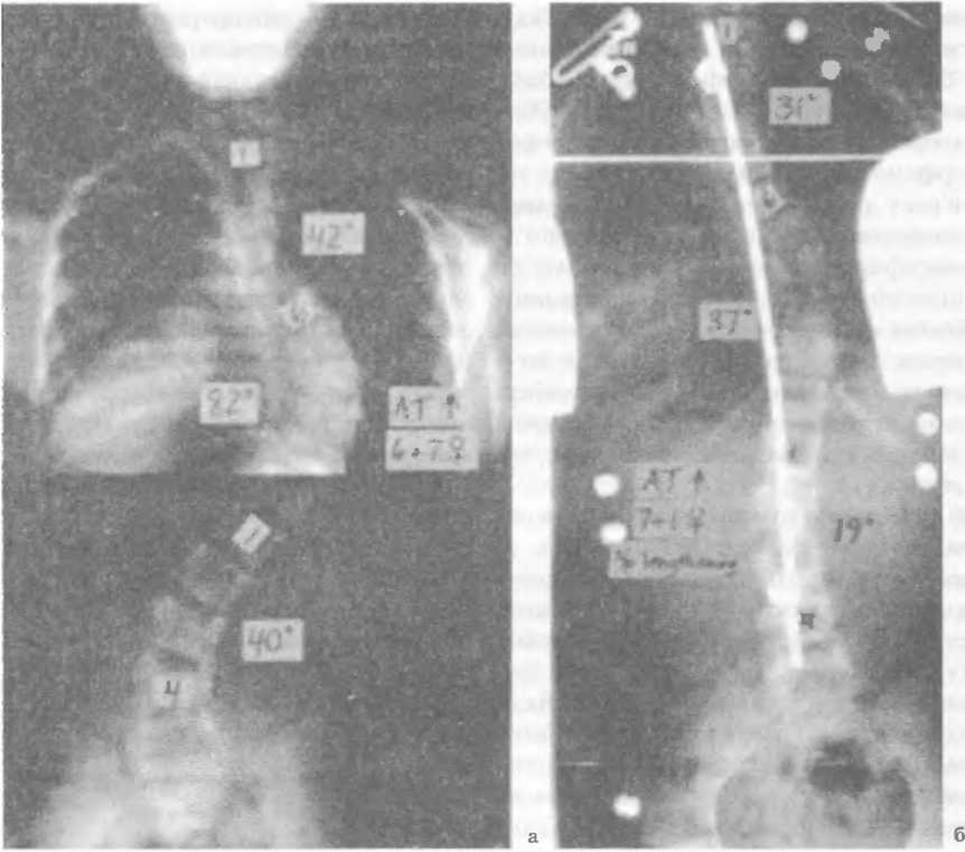
Fig. 70. Spinal deformity (Scoliosis) in Marfan Syndrome: a - before surgery, b - after corrective surgery by 37° and spinal fixation from Th2 to L5 (Hamill Ch.L., 1993).
Treatment is managed in the same manner as for degenerative-dystrophic processes.
Last update: 10/08/2026
Editorial and Educational Adaptation: This material has been compiled based on the primary/original source text. The project team performed an editorial review, corrected technical inaccuracies, structured sections, and adapted the content for an educational format.
What was processed:
- elimination of formatting defects (OCR errors, structural breaks, corrupted characters);
- editorial organization of content;
- standardization of terminology in accordance with academic sources;
- verification of factual statements against the original source text.
All mentions of the author, publication year, and origin of the primary text have been preserved in accordance with the source.