Orthopedics - Oleksa A.P. 2006
Kyphosis
Juvenile Kyphosis (Scheuermann's Disease)
The disease was first described in 1921 by Scheuermann. Spinal osteochondropathy is accompanied by Changes in the intervertebral discs, vertebral bodies, and their endplates, resulting in The Development of juvenile Kyphosis (Fig. 264).
The exact Etiology of the disease remains unclear to this day. As early as 1930, after examining the morphological changes in the spine, Schmorl ruled out a primary role of the apophyseal rings in the growth of vertebral bodies and the progression of the pathological process. In chondropathy, he observed alterations in the discs, the cartilaginous epiphyseal plate, and the Bone tissue of the vertebrae. As a result of these changes, elements of the cartilaginous plate and disc herniate into the cancellous bone of the vertebral body, forming a Schmorl's node.
Later theories emerged regarding the aseptic Necrosis of the apophyseal ring and abnormal endochondral ossification, which causes weakness of the Cartilage and plate, leading to anterior compression of the vertebral bodies under physical stress (Greene T.L. et al., 1985). Furthermore, the condition has been linked to genetic predisposition, inherited in an autosomal dominant manner (Lowe T.G., 1990), as well as dysontogenetic inferiority of the discs (Abalmasova E.A., Svintsov A.P., 1980; Abalmasova E.L. et al., 1984).
Class="center">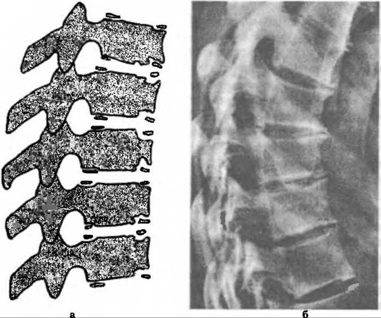
Fig. 264. Diagram (a) and radiograph (b) in Scheuermann's disease.
Clinically, the disease has an insidious onset and a slow progression, most commonly manifesting in boys around ten years of age.

Fig. 265. A patient with juvenile adolescent thoracic kyphosis.
Juvenile kyphosis manifests as a sagittal kyphotic curvature of the spine (Fig. 265), the apex of which is located in the region of the seventh to ninth thoracic vertebrae in the thoracic type, and in the region of the tenth thoracic and first lumbar vertebrae in the thoracolumbar type (Lowe T.G., 1990).
Clinically, the disease has a slow course divided into three stages.
The First stage presents as poor posture with a moderately increased kyphosis in the thoracic or thoracolumbar spine, which is stable and does not disappear when the patient lies flat on a hard surface. This stage persists until the Ossification of the vertebral body apophyses begins.
The Second Stage of the disease coincides with the appearance of the first ossification centers in the apophyses, at which point all characteristic clinical and radiological signs of kyphosis become established. Depending on the type, the spine is kyphotically curved and fixed in the thoracic or thoracolumbar region. In this stage, the spine tilts slightly to the side, clearly revealing Asymmetry of the shoulders and scapulae, as well as trunk inclination. Lateral curvature of the spine can be easily distinguished from Scoliosis by the absence of an S-shaped deformity and, radiographically, by the absence of vertebral rotation (torsion).
In this stage, only some children may experience pain in the region of the affected vertebrae, although Svintsov and Abalmasova note that pain occurs in 25% of outpatients and 52.5% of inpatients. These localized pains typically arise after physical exertion and subside with rest. Their onset is attributed to vertebral Osteoporosis and tension of the interspinous ligaments. Spinal mobility is almost always fully preserved, although the tension sign (Lasegue's sign) is occasionally positive.
Radiological findings include a downward shift of the physiological kyphosis, wedge-shaped deformity with uneven, jagged contours of the vertebral bodies, and anterior marginal protrusions (anterior marginal detachments, Lenke L.G., 1997). The endplates are sclerosed and fragmented. Schmorl's nodes are present, penetrating the vertebral bodies and surrounded by sclerotic rims. The intervertebral disc spaces in the affected area are narrowed. These radiological changes can vary in severity depending on the timing of the X-ray Examination and the course of the disease. The dynamics of radiological spinal changes also depend on the child's GROWTH AND DEVELOPMENT (Fig. 266).
In the Third Stage of juvenile kyphosis, the pathological process stabilizes, coinciding with the complete fusion of the apophyses with the vertebral bodies. The kyphosis is rigidly fixed, leading to a compensatory increase in the range of motion in the lordotic regions. This occasionally causes spinal instability and leads to radicular pain following physical exertion. The back Muscles are in an antalgic spasm.
Juvenile kyphosis must sometimes be differentiated from epiphyseal Dysplasia, Various Forms of spinal disorders of dysontogenetic origin (such as Güntz juvenile kyphosis, primary juvenile osteochondrosis, and Hindeman's fixed round back), among others.
Treatment for juvenile kyphosis must be comprehensive and exclusively conservative. The most crucial component is the regular execution of physical exercises aimed at hyperextension (reclination) of the kyphosis. Children are advised to Sleep on a firm mattress with a board, perform exercises several times a day, and undergo swimming, traction, back massage, thermal Procedures, and physio-balneotherapy. School-aged children are encouraged to wear a backpack rather than a single-strap bag. A rather effective device for maintaining posture and correcting kyphosis consists of upper straps secured over the shoulders and a lower belt around the torso. The device comprises two thin, elastic, non-conductive plates 20 mm wide, connected at the top and bottom by two fasteners leaving a 5 mm gap between their planes so they do not Touch. The lower fastener holds the plates rigidly, while the upper one can slide within a slot in the upper plate, retained by a pinhead. One plate is wired to the positive terminal and the other to the negative terminal of a pocket battery. In the middle of the upper plate, There is a thin 3-mm protrusion soldered in place and directed toward the underlying plate, without touching it when the plates are straight. When the plates are bent, the upper one approaches the underlying plate, making contact with the soldered protrusion, thereby closing the electrical circuit connected to an auditory alarm device.

Fig. 266. Spinal radiograph in juvenile osteochondropathic kyphosis.
Using this device, the child must constantly monitor their upright posture; otherwise, any increase in physiological kyphosis triggers an acoustic signal. Devices operating on a similar principle have been developed with a light signal, as well as mechanical ones where the protrusion soldered onto the upper plate is relatively sharp, so that spinal flexion causes it to pierce through an opening in the underlying plate and prick the child.
Strengthening the back and shoulder girdle muscles is essential. The Use of corrective braces that lead to Muscle atrophy should be avoided. Children are prescribed A balanced diet rich in Vitamins and Proteins. Spending summer vacations at the seaside while continuing treatment is strongly recommended.
Since patients with juvenile kyphosis require dynamic monitoring, they must be kept under dispensary medical surveillance.
Last update: 10/08/2026
Editorial and Educational Adaptation: This material has been compiled based on the primary/original source text. The project team performed an editorial review, corrected technical inaccuracies, structured sections, and adapted the content for an educational format.
What was processed:
- elimination of formatting defects (OCR errors, structural breaks, corrupted characters);
- editorial organization of content;
- standardization of terminology in accordance with academic sources;
- verification of factual statements against the original source text.
All mentions of the author, publication year, and origin of the primary text have been preserved in accordance with the source.