Medical Radiology - Lazar A.P. 2008
Radiation Therapy
Radiation Reactions and Injuries
During Radiation therapy, ionizing radiation acting on the patient's body Tissues can cause radiation reactions and injuries, the severity and duration of which depend on the dose and the fractionation schedule.
Inadequate control of dose distribution in the irradiated area may lead to regions with a high absorbed dose exceeding the tolerance limits of normal tissues, especially during repeated courses of radiation therapy. The Development of complications arising during postoperative radiation therapy is facilitated by Impaired Blood supply in the operated tissues and secondary infection.
The basis of radiation tissue injuries includes vascular obliteration, degenerative changes in nerve endings, and tissue atrophy, followed by replacement with coarse, unstructured Connective Tissue and the development of fibrosis (Fig. 331). Often, these changes are progressive and require specialized, prolonged Treatment.
In clinical practice, radiation complications are generally classified into radiation reactions and injuries based on their severity and the time it takes for them to resolve or diminish. Radiation reactions refer to temporary, self-limiting functional and organic disorders, whereas radiation injuries denote irreversible changes in human tissues and Organs caused by the effects of ionizing radiation.
Class="center">
Fig. 331. Radiation-induced fibrosis of the lower lobe of the right lung.
The Classification of radiation reactions and complications can be represented as follows:
I. Radiation reactions:
1. Local:
A. Skin: erythema, dry and moist epidermitis.
B. Mucous membranes: hyperemia, edema, mucositis, keratinization, and epithelial desquamation.
C. Internal Organs: laryngitis, esophagitis, pleuritis, pneumonitis, pneumosclerosis, myocarditis, pericarditis, gastritis, enterocolitis, proctitis, cystitis.
2. Systemic: disorders of organ or system function (headache, nausea, vomiting, tachy- or bradycardia, hypo- or Hypertension), Changes in the morphological COMPOSITION OF THE blood (anemia, leukopenia, thrombocytopenia).
II. Radiation injuries:
1. Local:
A. Skin: atrophy, indurative edema, radiation ulcer, radiation Cancer.
B. Mucous membranes: atrophy, radiation ulcer, fistulas, radiation cancer.
C. Internal organs: fibrosis, ulcers, necrosis, radiation cancer.
2. Systemic: persistent changes in the morphological composition of the blood, damage to organs and Organ Systems, chronic radiation sickness.
1. Radiation reactions.
General radiation reactions occur after irradiation of large areas of the body. They develop at the end of a course of radiation therapy and typically resolve spontaneously or with appropriate treatment. During radiation therapy, some patients may exhibit functional Disorders of the Central Nervous system, hematopoiesis, and the gastrointestinal tract. The General condition of patients is also affected by functional and morphological changes in the irradiated organs and tissues. This may manifest as headaches, drowsiness, polyuria, vasomotor instability, increased sweating, and irritability. Disturbances involving the cardiovascular, respiratory, and digestive systems may also be observed. These changes are predominantly functional in nature and tend to resolve after the completion of the course of radiation therapy. All these pathological processes in organs and systems gradually subside and return to normal within 3–8 months.
Identical doses elicit different responses in adults and children. Children's skin is more radiosensitive.
1. Erythema is reddening of the skin accompanied by Swelling, itching, a feeling of warmth, and pain. The underlying mechanism of erythema is a vascular reaction involving capillary dilation. Erythema occurs at a dose of 8 Gy with X-ray therapy and 30–35 Gy with gamma therapy. Two to three weeks after the completion of irradiation, erythema disappears and is replaced by pigmentation that persists for several months (Fig. 332).
2. Dry dermatitis (epidermitis) develops 2–3 weeks after irradiation, with cyanosis appearing against the Background of erythema and edema. The reaction culminates in the desquamation of the cornified layer of the epidermis, after which the skin becomes dry, pigmented, and atrophic. It develops at a dose of 25 Gy during X-ray therapy and 40–45 Gy during gamma therapy.
3. Moist dermatitis is characterized by hyperemia and skin edema, accompanied by blisters containing serous or purulent fluid. The blisters rupture, forming a weeping, bright pink surface that subsequently becomes covered with purulent crusts. Epithelialization begins beneath these crusts, after which the skin becomes dry, pigmented, and atrophic. It occurs at a dose of 30–35 Gy with X-ray therapy and 50–55 Gy with gamma therapy.
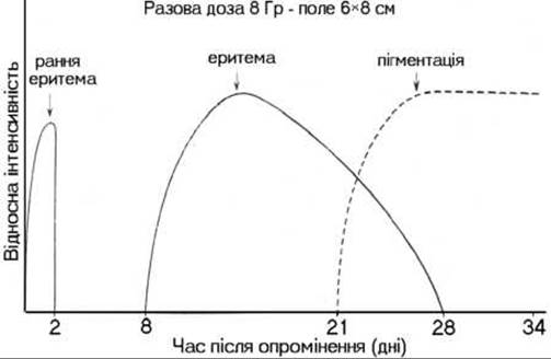
Fig. 332. Evolution of radiation reactions following exposure to a single dose of 8 Gy (according to Hellriegel).
All specified radiation skin reactions are accompanied by alopecia, which can be temporary or permanent. Following the resolution of the erythema reaction, Hair regrows within 2–3 months. After dry epidermitis, hair grows back, but often appears compromised—sparse, dry, and brittle. Occasionally, the same type of hair regrowth is observed after moist epidermitis as well.
Radiation Reactions of the mucous membranes are referred to as mucositis or radioepithelitis. Radioepithelitis is a progressive dystrophic process that develops through distinct stages. The First stage is characterized by mild hyperemia and mucosal edema. As the dose increases, these manifestations progress to epithelial keratinization. The Second Stage (focal mucositis) involves epithelial desquamation, where the keratinized epithelium detaches in certain areas, initially forming isolated erosions with a necrotic coating. In the Third Stage (confluent or membranous mucositis), significant shedding of the keratinized epithelium occurs alongside the coalescence of single erosions. In the Fourth Stage (erosive mucositis), against the background of extensive epithelial shedding, single or multiple erosions develop. The radiation reaction of the serous membrane concludes with the epithelialization of the erosions, leaving residual signs of edema and hyperemia.
Lately, with The Use of substantial radiation doses, radioepithelitis almost invariably accompanies radiation therapy for malignant tumors. With contact radiation therapy Methods (interstitial, intracavitary, or surface application), radioepithelitis develops in localized areas and reaches its maximum severity after the radiation source is removed. Mild hyperemia of the skin and mucous membrane is observed 30–36 hours after treating lower lip cancer with a dose of 20–30 Gy. By days 4–7, as the absorbed dose reaches 35–55 Gy, the radiation reaction intensifies. On days 15–21 following the removal of radioactive sources, the reactive symptoms subside, and epithelialization of the skin and mucous membranes begins.
Tab. 3. Gamma-ray tolerance doses providing a 5% probability of radiation injury to various organs and tissues.
|
Tissue |
Volume |
Dose (Gy/week) |
TDF Units |
|
Connective tissue |
<500 cm3 |
63/6 |
107 |
|
>500 cm3 |
60/6 |
100 |
|
|
Whole organ |
30/3 |
50 |
|
|
<50 % of organ |
40/4 |
66 |
|
|
Whole organ |
20/2 |
33 |
|
|
< 1/3 of organ |
60/6 |
100 |
|
|
Whole organ |
30/3 |
50 |
|
|
<100 cm3 |
60/6 |
100 |
|
|
Skin |
<2 cm (diameter) |
90/3 |
200 |
|
<10 cm (diameter) |
55/3 |
125 |
|
|
<30 cm (diameter) |
45/3 |
92 |
|
|
<5 cm (length) |
45/3 |
92 |
|
|
<10 cm |
50/5 |
82 |
|
|
>10 cm |
45/4,5 |
75 |
|
|
Intestines |
<100 cm3 |
45/4,5 |
75 |
|
Whole abdomen |
30/3 |
50 |
During radiation therapy for laryngeal tumors, the radiation reaction manifests as laryngitis of varying intensity. Along with radioepithelitis—which occurs at absorbed doses of 30–40 Gy during X-ray therapy and 50 to 55 Gy during gamma therapy—patients experience dry Mouth, sore throat, hoarseness, and a cough productive of abundant viscous sputum. Perichondritis develops if the perichondrium of the thyroid, cricoid, and arytenoid cartilages is compromised and becomes infected.
Radiation esophagitis occurs following radiotherapy for esophageal cancer and is accompanied by pain during swallowing of food and saliva, as well as a burning sensation in the Esophagus.
Radiation-induced pulmonary reactions are classified into: 1) acute reactions, resembling bronchopneumonia, which are reversible in most cases; 2) chronic reactions, resulting in irreversible changes such as pneumosclerosis and fibrosis.
2. Radiation injuries.
Radiation therapy for malignant tumors can damage surrounding healthy tissues, resulting in a characteristic presentation of local radiation injury. The frequency and severity of such injuries depend primarily on the cumulative absorbed dose of ionizing radiation (Table 3). Radiation injuries typically stem from errors in treatment planning and delivery, specifically when excessively high cumulative and single tumor doses are prescribed that exceed the tolerance limits of healthy tissues.
Depending on the time elapsed since the completion of radiation therapy, radiation injuries are categorized into early injuries, which develop within the first 3 months post-treatment, and late injuries, which manifest after 3 months. Organic and functional alterations in organs and tissues resulting from radiation injury require specialized medical management.
Last update: 08/08/2026
Editorial and Educational Adaptation: This material has been compiled based on the primary/original source text. The project team performed an editorial review, corrected technical inaccuracies, structured sections, and adapted the content for an educational format.
What was processed:
- elimination of formatting defects (OCR errors, structural breaks, corrupted characters);
- editorial organization of content;
- standardization of terminology in accordance with academic sources;
- verification of factual statements against the original source text.
All mentions of the author, publication year, and origin of the primary text have been preserved in accordance with the source.