Human Anatomy and Physiology - N. I. Fedyukovich 2003
Internal Organs
Digestive System
Oral Cavity
The Oral Cavity (cavitas oris) is the beginning of the Digestive System. With the help of the Teeth, food is crushed and chewed, while the Tongue softens and mixes it with saliva, which enters the oral cavity from the Salivary Glands, and then passes into the Pharynx.
The oral cavity is divided by the alveolar processes of the jaws and the teeth into two compartments: the Vestibule of the Mouth and the oral cavity proper.
The vestibule of the mouth (vestibulum oris) is a slit-like space bounded externally by the Lips and Cheeks, and internally by the upper and lower dental arches and Gums. The vestibule communicates with the external environment through the oral fissure, and with the oral cavity proper through the space formed by the upper and lower teeth and the gap behind the last molar. The oral fissure is bounded by the lips, which are musculocutaneous folds. The core of the lips is formed by the fibers of the orbicularis oris Muscle. In the corners of the mouth, the lips are joined by the labial commissures. The outer surface of the lips is covered with Skin, and the inner surface is lined with mucous membrane and stratified squamous non-keratinized epithelium. At the transition of the mucous membrane to the gums, the frenula of the upper and lower lips are located.
The oral cavity proper (cavitas oris propria) extends from the teeth to the entrance of the pharynx. It is bounded superiorly by the Hard and Soft palate, inferiorly by the Muscles that form the floor of the mouth, anteriorly and laterally by the cheeks and teeth, and posteriorly, through a wide opening, by the fauces.
The cheeks (buccae) are formed by the buccinator muscles. They are covered externally by skin and internally by mucous membrane. Between the skin and the buccinator muscles lies a thick layer of adipose tissue, which forms the buccal fat pad. It is particularly well developed in infants, facilitating the act of sucking. The duct of the Cytology/practical/97.html">Parotid salivary gland opens on the mucous membrane of the cheek, within the vestibule of the mouth.
The gums (gingivae) are a continuation of the mucous membrane of the lips and cheeks; they cover the alveolar processes of the jaws and tightly envelop the necks of the teeth.
The tongue (lingua) is a muscular organ involved in mixing food in the oral cavity, taste perception, swallowing, and articulation. The tongue is located on the floor (lower wall) of the oral cavity. It is a flat, elongated oval body. The tongue has an apex, a body, and a ROOT, as well as an upper surface (dorsum of the tongue), a lower surface, and margins. The mucous membrane of the dorsum of the tongue forms projections called papillae of various SHAPES AND SIZES. These include fungiform, foliate, filiform, conical, and vallate papillae. They contain Blood Vessels and nerve endings for taste or general sensation. The mucous membrane of the root of the tongue has no papillae. Instead, it contains numerous lymphoid nodules that form the lingual tonsil. On the lower surface of the tongue, as the mucous membrane reflects onto the floor of the mouth, it forms a midline fold—the frenulum of the tongue.
The Muscles of the tongue (m. linguae) are paired and divided into extrinsic and intrinsic muscles. The extrinsic muscles include three muscles: the genioglossus (m. genioglossus), which protrudes the tongue forward or deviates it to the side; the hyoglossus (m. hyoglossus), which depresses and retracts the tongue; and the styloglossus (m. styloglossus), which elevates and retracts the tongue. The intrinsic muscles of the tongue are represented by four muscles that run within the body of the tongue and intersect in mutually perpendicular directions: the superior and inferior longitudinal muscles, the transverse, and the vertical muscles. Upon contraction, they alter the shape of the tongue.
Teeth (dentes) are located in the dental alveoli of the upper and lower jaws along the upper margin of the gums. Teeth serve as Organs for grasping, biting, and grinding food, and they also participate in speech production.
In humans, teeth are replaced twice during their lifetime: first, 20 deciduous teeth appear in a specific sequence, followed by 32 permanent teeth. All teeth have a similar Structure. Each tooth consists of a crown, a neck, and a root. The crown is the most massive part of the tooth, projecting above the gum. It has lingual, vestibular (facial), contact, and occlusal (chewing) surfaces.
By means of a special type of continuous joint—gomphosis—the teeth are immovably fixed in the dental alveoli of the jaws. Each tooth has one to three roots. The root ends in an apex, which has a small apical foramen through which Vessels and nerves enter and exit the pulp cavity. The root is held in the dental alveolus of the jaw by Connective Tissue—the periodontium. The neck of the tooth is a slight constriction between the crown and the root, enveloped by the mucous membrane of the gum. Inside the tooth, There is a small pulp cavity, which forms the crown cavity and continues into the root as the root canal. The pulp cavity is filled with dental pulp, consisting of connective tissue, blood vessels, and nerves. The dental Tissues include dentin, enamel, and cementum. Dentin surrounds the pulp cavity and root canal, forming the bulk of the tooth. Externally, the crown is covered with enamel, and the root with cementum.
The teeth of an adult are arranged symmetrically on the upper and lower jaws, with 16 teeth on each. They can be written as a formula:
Class="center">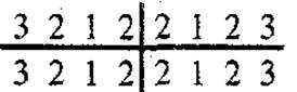
(2 incisors, 1 canine, 2 premolars, and 3 molars in each half).
Each tooth has its own shape and performs a corresponding function; for example, incisors are designed for cutting (shearing) food, canines for tearing, and molars for crushing and grinding.
The deciduous tooth formula is as follows:

The first deciduous teeth begin to erupt in infants at 5 to 7 months of age, and the process is completed by the beginning of the third year; they function only until 6 to 7 years of age. Then, before the eruption of the corresponding permanent tooth, the deciduous tooth falls out. Permanent teeth appear in children at the age of 6 to 7 years, and this process is completed by 13 to 15 years.
The palate (palatum) is divided into the hard and soft palate. The hard palate is formed by the palatine processes of the Maxilla and the horizontal plates of the palatine bones, joined together by the palatine suture. It is covered with a mucous membrane lined with stratified squamous non-keratinized epithelium, which is firmly fused to the periosteum.
The soft palate is a fibromuscular structure covered with mucous membrane. The anterior part of the soft palate lies horizontally, while the posterior part hangs freely, forming the palatine velum with the uvula in the middle. They separate the nasopharynx from the oropharynx. Extending from the lateral margins of the palatine velum are two folds (arches): the anterior palatoglossal arch and the posterior palatopharyngeal arch. The former descends to the lateral surface of the tongue, and the latter to the lateral wall of the pharynx. Between these arches lies the tonsillar fossa containing the palatine tonsil. The core of the soft palate is composed of paired skeletal muscles (tensor veli palatini, levator veli palatini, palatoglossus, and palatopharyngeus muscles) and the unpaired musculus uvulae. Upon contraction, they tense the palatine velum, elevate, and depress the soft palate.
Posteriorly, the oral cavity communicates with the pharynx through the isthmus of fauces. The isthmus of fauces is bounded superiorly by the soft palate, inferiorly by the root of the tongue, and laterally by the palatoglossal arches.
Last update: 08/08/2026
Editorial and Educational Adaptation: This material has been compiled based on the primary/original source text. The project team performed an editorial review, corrected technical inaccuracies, structured sections, and adapted the content for an educational format.
What was processed:
- elimination of formatting defects (OCR errors, structural breaks, corrupted characters);
- editorial organization of content;
- standardization of terminology in accordance with academic sources;
- verification of factual statements against the original source text.
All mentions of the author, publication year, and origin of the primary text have been preserved in accordance with the source.