Human Anatomy, Part 1 - K. A. Dyubenko, A. K. Kolomiyytsev, Yu. B. Chaykovsky 2002
Special Part
Splanchnology, splanchnologia [the study of viscera] — Development of the Digestive and Respiratory Systems
Development of the Face, Oral, and Nasal Cavities
At the end of the first month of development, two ectodermal depressions form On the surface of the embryo's body, directed towards the gut tube. The anterior depression, the stomodeum (primary Oral Cavity), is located on the ventral surface of the HEAD and, during this period, is pressed against the chest in the area of the future oral cavity. The posterior depression, the proctodeum, is situated more caudally than the future anal region. As the floor of the stomodeum deepens, it comes into contact with the lining of the foregut, resulting in The formation of the buccopharyngeal membrane. During the fourth week, this membrane establishes the transition point from the oral cavity to the Pharynx. Initially, the stomodeal depression is shallow, but later, due to the growth of its surrounding structures, a deep oral cavity is formed.
The stomodeal depression is bounded superiorly by a prominent, overhanging frontonasal process. This process incorporates the expanded cranial end of the neural tube, which serves as the primordium of the Brain. Inferiorly, the stomodeum is bounded by the first branchial (mandibular) arch (Fig. 274A). Subsequently, two nasal pits appear within the upper frontonasal process, each surrounded by medial and lateral elevations, known as the medial and lateral nasal processes. The mandibular arch is subdivided by grooves into paired maxillary processes—located inferior to the lateral nasal processes—and paired mandibular processes (Fig. 274B). Thus, the opening of the primary oral cavity is surrounded by five prominences: superiorly by the unpaired frontonasal process, and laterally by the paired maxillary and mandibular processes. Between the lateral nasal and maxillary processes, naso-optic furrows develop, extending from the site of eye formation to the nasal pits. A small triangular area forms between the medial nasal processes, which later contributes to the Formation of the nasal bridge and alae.
During the sixth week, the maxillary processes experience a marked increase in volume, growing towards the midline and bringing the nasal processes closer together. The medial nasal processes shift toward the midline, fusing with each other and with the maxillary processes to form the upper jaw. The mandibular processes also fuse along the midline to form the lower jaw.
The accelerated growth of the structures surrounding the stomodeum drives the formation of the external facial features, jaws, and oral cavity walls. This process is accompanied by the medial shift of the eyes from their initial lateral position (Fig. 274D, E). The groove separating the maxillary and lateral nasal processes transforms into the nasolacrimal canal, canalis nasolacrimalis, which connects the future orbital cavities with the future Nasal cavity.
By the end of the second month of development, with the formation of the upper jaw, palatal processes emerge. As with the upper jaw, these processes develop from the maxillary and medial nasal processes, with the greater part of the palate being formed by the maxillary processes. Outgrowths appear on the inner surfaces of these processes and expand toward the midline. Since the Tongue initially occupies the space between them, these outgrowths are directed downward, with their edges lying lateral to the tongue. As the tongue shifts downward, the edges of the palatal processes elevate slightly and fuse along the midline. The formation of the palate divides the stomodeal cavity into oral and nasal cavities. Concurrently with palatogenesis, the nasal septum grows toward the palate, extending backward and downward from the frontonasal process and connecting with its superior surface. This results in the division of the nasal cavity into left and right halves.
Class="center">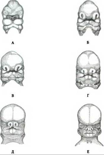
Fig. 274. Key stages of facial morphogenesis in Embryology (after Patten):
A - 4-week embryo; B - 5-week embryo; C - 5.5-week embryo; D - 6-week embryo; E - 7-week embryo; F - 7.5-week embryo;
A. 1 - frontonasal process; 2 - nasal placode; 3 - buccopharyngeal membrane; 4 - maxillary process; 5 - mandibular arch; 6 - hyoid arch.
B. 1 - frontonasal process; 2 - nasal pit; 3 - oral opening; 4 - maxillary process; 5 - mandibular arch; 6 - hyoid arch.
C, D. 1 - medial nasal process; 2 - lateral nasal process; 3 - nasohyal groove;
4 - maxillary process; 5 - lower jaw; 6 - hyomandibular cleft.
E. 1 - lateral nasal process; 2 - medial nasal process; 3 - auricular hillocks surrounding the hyomandibular cleft; 4 - Hyoid bone; 5 - laryngeal Cartilage.
F. 1 - lateral nasal process; 2 - fused medial nasal processes (forming a midline groove in the upper lip); 3 - External ear; 4 - hyoid bone; 5 - laryngeal cartilages
The formation of the Nose is associated with the medial migration of the nasal pits and the lateral and medial processes bounding them. The medial processes unite to form the central part of the upper lip and the nasal septum. The alae of the nose are formed by the lateral nasal processes.
During these changes, the nasal pits deepen and transform into passages that open into the primary oral cavity. The external openings of these channels become the nostrils, while the internal openings empty into the oral cavity to form the choanae, between which lies the aforementioned primordium of the nasal septum.
The primary oral cavity opens externally via the primary oral orifice. Its dimensions gradually change as the margins grow and the Cheeks develop (Fig. 274D). The epithelium along the edges of the oral opening thickens, resulting in the formation of the labiodental lamina, within which a groove appears that gives rise to the Vestibule of the Mouth, vestibulum oris.
Various anomalies may arise in the facial region during development (Fig. 275), the most common being congenital cleft lip (popularly known as "harelip") and cleft palate (cleft of the upper lip, jaw, and hard palate). According to L. V. Kharkov (1998), various combinations of these anomalies can occur: unilateral cleft of the upper lip, alveolar process, and Hard and Soft palate; congenital symmetric bilateral cleft of the upper lip, alveolar process, and hard and soft palate; congenital right-sided macrostomia, occult right-sided coloboma (Greek *coloboma* — a missing part of the eyelid margin), and others.
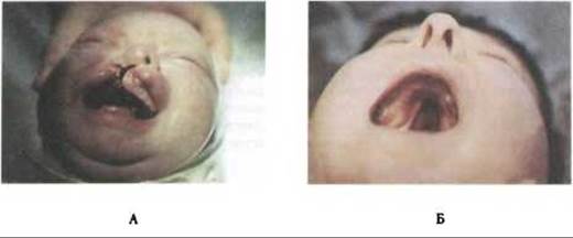
Fig. 275. Most frequent Developmental anomalies of the face and palate (after L. V. Kharkov):
A - unilateral cleft of the upper lip, alveolar process, and hard and soft palate; B - isolated congenital cleft of the soft palate and 2/3 of the hard palate
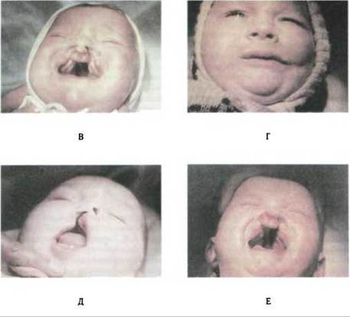
Fig. 275. Most frequent developmental Anomalies of the face and palate (after L. V. Kharkov) (Continued):
C - congenital symmetric bilateral cleft of the upper lip, alveolar process, hard and soft palate; D - right-sided oblique facial cleft; E - unilateral congenital cleft of the upper lip, alveolar process, and palate; k - right-sided congenital cleft of the upper lip and alveolar process; F - congenital bilateral cleft of the upper lip, alveolar process, and hard and soft palate with protrusion of the premaxilla
Last update: 08/08/2026
Editorial and Educational Adaptation: This material has been compiled based on the primary/original source text. The project team performed an editorial review, corrected technical inaccuracies, structured sections, and adapted the content for an educational format.
What was processed:
- elimination of formatting defects (OCR errors, structural breaks, corrupted characters);
- editorial organization of content;
- standardization of terminology in accordance with academic sources;
- verification of factual statements against the original source text.
All mentions of the author, publication year, and origin of the primary text have been preserved in accordance with the source.