MEDICAL BIOLOGY, HUMAN ANATOMY, PHYSIOLOGY AND PATHOLOGY - Ya.I. Fedoniuk 2010
ANATOMY, PHYSIOLOGY, PATHOLOGY
SECTION 3. ANATOMICAL AND PHYSIOLOGICAL ASPECTS OF THE SELF-REGULATION OF BODY FUNCTIONS
MENINGES
The Meninges of the Brain are a continuation of the spinal meninges. The dura mater encephali differs from the spinal dura mater in the following features:
1. It serves as the periosteum of the cranial bones, meaning there is no epidural space here.
2. In certain areas, it forms a series of folds that project between PARTS OF THE brain—specifically, the falx cerebri, falx cerebelli, tentorium cerebelli, and diaphragma sellae (Fig. 3.35).
Class="center">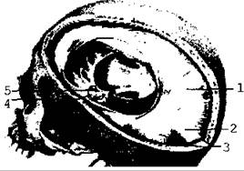
Fig. 3.35. Dura mater of the brain
3. These folds split and attach to the margins of the sulci on the cranial bones, forming venous sinuses filled with venous Blood. Venous blood from the sinuses drains into the Internal jugular vein (Fig. 3.36).
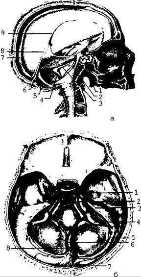
Fig. 3.36. Dural Venous Sinuses: a - lateral view; b - superior view
The arachnoid mater (arachnoidea encephali) is thin, transparent, and vascular. It bridges across the sulci and fissures on the brain's surface. In the region of these depressions, enlargements of the subarachnoid space called cisterns are formed. They are filled with CEREBROSPINAL FLUID. The largest cisterns are located between the Cerebellum and the Medulla Oblongata, as well as at the Base of the brain.
The arachnoid mater forms small projections (arachnoid granulations, or Pacchionian bodies) that penetrate the venous sinuses, allowing cerebrospinal fluid to drain into the bloodstream.
The pia mater (pia mater encephali) is vascularized and extends into all the fissures and sulci of the brain. It participates in The formation of the choroid plexuses of the ventricles.
The intermeningeal spaces are identical to those surrounding the Spinal Cord, with the exception of the epidural space (Fig. 3.37).
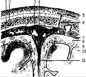
Fig. 3.37. Schematic representation of the relationships between the meninges, the superior sagittal sinus, the cranial vault, and the brain surface in a frontal section
Last update: 08/08/2026
Editorial and Educational Adaptation: This material has been compiled based on the primary/original source text. The project team performed an editorial review, corrected technical inaccuracies, structured sections, and adapted the content for an educational format.
What was processed:
- elimination of formatting defects (OCR errors, structural breaks, corrupted characters);
- editorial organization of content;
- standardization of terminology in accordance with academic sources;
- verification of factual statements against the original source text.
All mentions of the author, publication year, and origin of the primary text have been preserved in accordance with the source.