MEDICAL BIOLOGY, HUMAN ANATOMY, PHYSIOLOGY AND PATHOLOGY - Y.I. Fedoniuk 2010
ANATOMY, PHYSIOLOGY, PATHOLOGY
CHAPTER 1. ORGAN. ORGAN SYSTEM. ORGANISM
TUMORS
CLASSIFICATION OF TUMORS
3. MESENCHYMAL TUMORS
3.1. SOFT TISSUE TUMORS
Benign mesenchymal tumors. Fibroma is the most common tumor originating from mature Fibrous Connective Tissue. It can occur in any organ, but is most frequently found in the Skin, mammary gland, and Uterus. Fibroma is characterized by tissue atypia, manifested by a chaotic arrangement of connective tissue fibers and an uneven distribution of Blood Vessels. The tumor grows expansively and possesses a capsule. The Clinical significance of a fibroma depends on its localization: a cutaneous fibroma causes little concern to the patient, whereas a fibroma within the spinal canal can cause severe neurological impairments.
Lipoma is a tumor of mature adipose tissue that grows expansively as single or multiple nodes and typically has a capsule. It is most commonly located in the subcutaneous adipose tissue. Occasionally, a lipoma can reach considerable size.
Myoma is a tumor derived from Muscle tissue. A distinction is made between leiomyoma (originating from smooth muscle) and rhabdomyoma (originating from striated muscle). Tissue atypia consists of varying thicknesses of muscle bundles running in different directions and forming whorls. Tumors in which the stroma predominates are referred to as fibromyomas. Leiomyomas are most frequently encountered in the uterus, where they sometimes attain significant dimensions. Rhabdomyoma is a rarer tumor that may arise in the Muscles of the Tongue, myocardium, and other Organs containing Striated muscle tissue.
Hemangiomas are a group of vascular tumors. Depending on the type of blood vessels giving rise to tumor growth, capillary, venous, and cavernous hemangiomas are distinguished. Capillary hemangioma is generally congenital and localized in the skin as red-brown patches with an irregular surface. Venous angioma consists of vascular cavities resembling Veins. Cavernous hemangioma is composed of vascular channels of varying size and shape with walls of unequal thickness. Thrombi frequently form within these vascular spaces. Upon trauma, a cavernous hemangioma can cause significant Hemorrhage. Venous and cavernous angiomas are most commonly found in the Liver, muscles, and occasionally in the bones and Brain.
Malignant mesenchymal tumors. These tumors are collectively called sarcomas because on cross-section they resemble fish meat (from the Greek sarcos - flesh). They are accompanied by tissue and cellular atypia, as well as hematogenous metastasis, As a result of which metastases appear quite early and become widespread. For these reasons, sarcomas exhibit a highly malignant clinical course. They develop from the same Tissues (mesenchymal derivatives) as benign mesenchymal tumors. Several types of soft tissue sarcomas are distinguished.
Fibrosarcoma arises from fibrous connective tissue, appears as a node with indistinct borders, infiltrates surrounding tissues, and is characterized by a fairly malignant course.
Liposarcoma develops from immature fat Cells. It can reach large sizes and remain free of metastases for a long time.
Myosarcomas, depending on the type of muscle tissue, are divided into leiomyosarcomas (Fig. 1.5), which develop from Cytology/cytology/32.html">Smooth muscle tissue, and rhabdomyosarcomas, which develop from striated muscles (Fig. 1.6). The cells of these tumors are characterized by atypia and polymorphism, often completely losing resemblance to muscle tissue, which makes identification of the original tissue possible only with the aid of an Electron microscope.
Angiosarcoma is a malignant vascular tumor. It has an exceptionally aggressive clinical course and gives rise to early hematogenous metastases.
Class="center">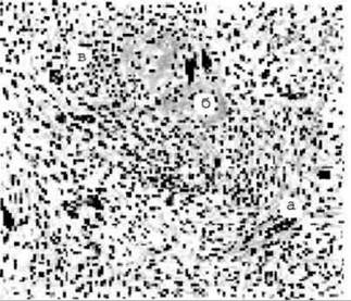
Fig. 1.5. Leiomyosarcoma: smooth muscle cells of various shapes; atypical cells with irregularly shaped nuclei are visible among them (a). The upper part of the image shows an area of necrosis (b) and an accumulation of leukocytes (c). Hematoxylin and eosin staining.

Fig. 1.6. Rhabdomyosarcoma: among the polymorphic tumor elements, giant atypical cells with granular Cytoplasm (a) and elongated cells with cross-striations (b) are visible. Hematoxylin and eosin staining.
Last update: 08/08/2026
Editorial and Educational Adaptation: This material has been compiled based on the primary/original source text. The project team performed an editorial review, corrected technical inaccuracies, structured sections, and adapted the content for an educational format.
What was processed:
- elimination of formatting defects (OCR errors, structural breaks, corrupted characters);
- editorial organization of content;
- standardization of terminology in accordance with academic sources;
- verification of factual statements against the original source text.
All mentions of the author, publication year, and origin of the primary text have been preserved in accordance with the source.