MEDICAL BIOLOGY, HUMAN ANATOMY, PHYSIOLOGY AND PATHOLOGY - Ya.I. Fedonyuk 2010
ANATOMY, PHYSIOLOGY, PATHOLOGY
CHAPTER 1. ORGAN. ORGAN SYSTEM. ORGANISM
INFLAMMATION
2. CLASSIFICATION OF INFLAMMATION
Depending on The Nature of the dominant local process (alteration, exudation, or proliferation), Three types of inflammation are distinguished: alterative, exudative, and proliferative (productive).
Alterative inflammation is dominated by tissue damage, such as dystrophy and necrosis. It occurs primarily in parenchymal Organs during infectious diseases accompanied by pronounced intoxication (e.g., caseous Necrosis of the Adrenal Glands or Lungs in tuberculosis patients).
Exudative inflammation is characterized by significant Circulatory Disorders accompanied by exudation and leukocyte emigration. Based on the Nature of the exudate, the following types are distinguished:
1. Serous inflammation.
2. Fibrinous inflammation.
3. Purulent inflammation.
4. Catarrhal inflammation.
5. Hemorrhagic inflammation.
6. Putrid inflammation.
7. Mixed forms.
Serous inflammation is characterized by The formation of a serous exudate containing up to 2% protein and a small number of cellular elements. This exudate can diffuse into the tissue of an organ, leading to inflammatory edema, accumulate in various Body Cavities (pleural, pericardial, peritoneal), or appear On the surface of mucous membranes. Causes of serous inflammation include thermal factors (burn disease), infectious agents (Mycobacterium tuberculosis, diplococci, meningococci), and autointoxication. Serous inflammation most commonly has an acute course. From the body's perspective, it is a favorable and mild form of the inflammatory process. Occasionally, serous inflammation in Internal Organs can lead to The Development of sclerosis.
Fibrinous inflammation is characterized by the formation of an exudate that tends to coagulate due to fibrin deposition. Depending on the depth of necrosis, Croupous and diphtheritic forms of inflammation are distinguished.
Croupous inflammation occurs when necrosis affects only the superficial layers of Tissues or membranes. It is most commonly observed on serous membranes (Pleurisy, pericarditis), mucous membranes—especially of the respiratory tract (tracheitis, Bronchitis)—and in the lungs (Croupous Pneumonia).
Macroscopically, the exudate on serous and mucous membranes appears as a grayish-white film that is easily detached; sometimes it can obstruct the lumen of organs such as the Trachea or Bronchi. The tissue surface in the inflamed area is hyperemic and dull.
Diphtheritic inflammation is distinguished by deeper necrosis that affects not only the surface layers but also penetrates deep into the tissues. The necrotic areas are infiltrated by a fibrinous exudate, forming a tough film firmly adherent to the underlying inflamed tissues. When this film is peeled away, ulcers are formed (Fig. 1.4).
Class="center">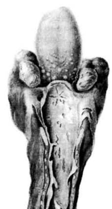
Fig. 1.4. Diphtheritic inflammation: diphtheritic tonsillitis, croupous laryngitis, and tracheitis.
Purulent inflammation is characterized by the formation of a purulent exudate consisting of A large number of leukocytes, protein, necrotic tissue debris, as well as living and dead microorganisms.
Purulent exudate, or pus, is a thick, yellow-green mass with an unpleasant odor. Purulent inflammation can localize on mucous membranes (purulent bronchitis, urethritis), serous membranes (purulent pleurisy, Empyema of the Pleura, pericarditis), and within internal organs (myocarditis, hepatitis, nephritis).
Purulent inflammation may have an acute, subacute, or chronic course. It is caused by pyogenic microorganisms, including various cocci, Pseudomonas aeruginosa, Escherichia coli, and certain rickettsiae.
At the same time, aseptic purulent inflammation can develop without the involvement of microorganisms, for instance, when certain substances (such as turpentine) are introduced into tissues.
The Significance of purulent inflammation for the body depends on its localization, spread, and complications. If an abscess is located in the Brain or Heart tissue, it poses an extreme, life-threatening danger. Should pus break into the bloodstream and microbes spread throughout the body against the Background of specific alterations in reactivity, Sepsis may develop. Nevertheless, timely Treatment—including surgical intervention—accelerates patient recovery.
Catarrhal inflammation is a condition in which exudate accumulating on The surface of mucous membranes flows off them, mixed with mucus produced by mucous glands. This mucus imparts viscosity to the exudate, which also contains leukocytes and desquamated epithelium. In this state, the mucous membrane appears thickened and edematous.
Catarrhal inflammation can be serous, mucous, or purulent.
It may also assume a chronic course (manifesting as hypertrophic and atrophic catarrhs).
Hemorrhagic inflammation develops when the exudate contains a significant number of erythrocytes, being characteristic of conditions such as anthrax, plague, and Influenza.
Putrescent, or ichorous, inflammation results from the invasion of putrefactive microflora into a focus of purulent inflammation, leading to tissue necrosis. As a rule, this type of inflammation is observed in debilitated patients with extensive, non-healing wounds or chronic abscesses. Morphologically, it is characterized by progressive tissue necrosis without a tendency toward demarcation. Necrotic tissues emit a foul odor, systemic intoxication increases, and the process frequently culminates in patient mortality.
Acute exudative inflammations frequently present a mixed character (e.g., purulent-fibrinous).
Productive (proliferative) inflammation is characterized by the proliferation of cellular and tissue elements, resulting in the development of focal and diffuse cellular infiltrates. Very little exudate is formed.
The following types of productive inflammation are distinguished:
- interstitial;
- granulomatous;
- hyperplastic proliferations of inflammatory origin.
In interstitial inflammation, the cellular infiltrate is localized within the interstitium of internal organs. A prolonged process leads to sclerosis, driven by Connective Tissue proliferation accompanied by signs of parenchymal atrophy (such as Liver cirrhosis or cardiosclerosis).
Granulomatous inflammation is based on the formation of a nodule (granuloma) in tissues as a manifestation of Cell proliferation and transformation.
Granulomatous inflammation underlies granulomatous diseases. Most commonly, these are specific granulomas of infectious origin, characteristic of tuberculosis, Syphilis, leprosy, rhinoscleroma, and glanders.
Nonspecific granulomas lack distinct diagnostic features. They occur in acute infectious diseases (such as epidemic or typhoid fever) as well as non-infectious conditions (silicosis, asbestosis, foreign body granulomas).
Hyperplastic proliferations of inflammatory origin are observed on mucous membranes and adjacent squamous epithelium. This condition is characterized by the formation of papillary outgrowths known as polyps (on mucous membranes) or condylomata acuminata (on transitional epithelium).
Last update: 08/08/2026
Editorial and Educational Adaptation: This material has been compiled based on the primary/original source text. The project team performed an editorial review, corrected technical inaccuracies, structured sections, and adapted the content for an educational format.
What was processed:
- elimination of formatting defects (OCR errors, structural breaks, corrupted characters);
- editorial organization of content;
- standardization of terminology in accordance with academic sources;
- verification of factual statements against the original source text.
All mentions of the author, publication year, and origin of the primary text have been preserved in accordance with the source.