MEDICAL BIOLOGY, HUMAN ANATOMY, PHYSIOLOGY AND PATHOLOGY - Ya.I. Fedoniuk 2010
ANATOMY, PHYSIOLOGY, PATHOLOGY
CHAPTER 1. ORGAN. SYSTEM OF ORGANS. ORGANISM
CIRCULATORY DISORDERS
5. INFARCTION
An infarction is a vascular (ischemic) necrosis resulting from an acute disruption of Blood flow. Infarctions typically occur in Internal Organs. Depending on their macroscopic appearance, Three types of infarctions are distinguished: white (ischemic), red (hemorrhagic), and mixed.
In a white (ischemic) infarction, the necrotic tissue is not saturated with blood. This occurs because, following the cessation of arterial blood inflow caused by the occlusion of an arterial branch, a reflex vasospasm develops both in the area supplied by the blocked artery and in the adjacent regions.
Such infarctions are most commonly found in the Spleen and Kidneys (Fig. 1.2 a, g, d).
A hemorrhagic infarction usually develops under conditions of venous congestion, for example, in the Lungs during Heart Failure, or in the spleen during thrombosis of its Veins. Drainage of blood from the area deprived of arterial supply fails to occur; moreover, blood accumulates excessively in this area due to influx through capillary anastomoses from adjacent regions. This leads to blood stasis accompanied by diapedetic hemorrhages (Fig. 1.2 b, c).
Class="center">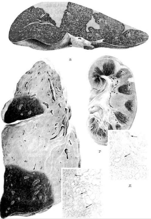
Fig. 1.2. Infarction: a - white (ischemic) infarctions of the spleen (indicated by arrows); b - red (hemorrhagic) infarctions of the lung (indicated by arrows); c - microscopic view of a Hemorrhagic Infarction of the lung: the necrotic zone is infiltrated with blood (indicated by arrows), hematoxylin and eosin staining; g - ischemic infarctions of the Kidney (indicated by arrows); d - microscopic view of an ischemic infarction of the kidney: the necrosis zone (lower arrow) is demarcated from the preserved tissue by a zone of marked hyperemia and leukocyte infiltration (upper arrow), hematoxylin and eosin staining.
A mixed infarction is also known as an ischemic infarction with a hemorrhagic rim. It develops against the Background of venous congestion in an organ typically prone to white infarction, such as the kidney. In this case, in the peripheral tissue surrounding the infarction, the initial reflex vasospasm is rapidly replaced by vasodilation, engorgement of vessel lumens, stasis, and diapedetic hemorrhages.
The outcomes of an infarction vary depending on its localization, size, the body's reactive capabilities, etc. It typically results in Organization, meaning The formation of a Connective Tissue scar at the site of the lesion. Infarction of the Brain results in cyst formation. Secondary infection leads to suppurative inflammation of the infarction, which in some cases can progress to gangrene; for instance, intestinal infarctions, which are constantly exposed to bowel contents, may result in putrefactive tissue breakdown in the form of wet gangrene.
Last update: 08/08/2026
Editorial and Educational Adaptation: This material has been compiled based on the primary/original source text. The project team performed an editorial review, corrected technical inaccuracies, structured sections, and adapted the content for an educational format.
What was processed:
- elimination of formatting defects (OCR errors, structural breaks, corrupted characters);
- editorial organization of content;
- standardization of terminology in accordance with academic sources;
- verification of factual statements against the original source text.
All mentions of the author, publication year, and origin of the primary text have been preserved in accordance with the source.