PLANT HETEROPHYLLY - O.M. NEDUKHA - 2011
CHAPTER ONE. MORPHOLOGICAL AND STRUCTURAL-FUNCTIONAL CHARACTERISTICS OF VEGETATIVE ORGANS IN HETEROPHYLLOUS PLANTS
1.7. LEAF EPIDERMIS IN HETEROPHYLLOUS PLANTS
1.7.3. Hydathodes, trichomes, and hydropotes
Hydathodes. The floating leaves of certain hydrophyte species and the basal rosette leaves of Campanula rotundifolia are characterized by specialized cellular structures known as hydathodes, which are responsible for the exudation of Water and aqueous solutions containing organic and inorganic substances (Takeda et al., 1991; Pederson, Sand-Jensen, 1993; Barabanov, 2006). Hydathodes consist of an epithem composed of chlorophyll-free epidermal Cells. The Structure of hydathodes in young leaves of Sparganium emersum (L.) Rehman has been described in detail: leaf hydathodes form a channel situated between two adjacent epidermal cells. The inner opening of the hydathode faces the xylem cells of vascular bundles and parenchymal cells, whereas the outer channel is surrounded by the lignified Cell walls of epidermal cells (Chen, Chen, 2005). The pecto-cellulosic walls of the two epidermal cells seal the hydathode opening. This aperture can remain open for extended periods; consequently, Algae and Bacteria entering the hydathode cavity form a "plug" near the open xylem element through which water enters. Such a plug may also consist of cutin, suberin, or callose (Pederson et al., 1997). The authors concluded that the minimum ROOT pressure driving water flow through the hydathodes of Sparganium emersum was 2.13 mL leaf-1 h-1, with a root pressure of approximately 3 kPa. In contrast, the root pressure in the hydrophyte Myriophyllum sp. was an order of magnitude higher, reaching 30 kPa (Pederson, 1994). It was established that hydraulic conductance (through hydathodes) at the apices of young leaves was higher than in older leaves, indicating an ontogenetic dependence of leaf hydraulic conductance in the studied hydrophyte. Guttation in young leaves of Fragaria L. (Takeda et al., 1991) and Lobelia L. (Pederson, Sand-Jensen, 1993) has been described previously.
Trichomes. The presence of trichomes has been observed On the surface of floating leaves of Trapa natans (Bercu, 2004) and Victoria amazonica Sowerby (Carpenter, 2006), as well as aerial leaves of Marsilea quadrifolia (Lin et al., 2007; Tai-Chung Wu, 2010). Structurally, these trichomes are similar to those found on the leaves of terrestrial plants (Werker, 2000). A trichome is a unicellular or multicellular structure on the leaf epidermal surface (Fig. 1.7.3.1, d) through which various metabolites and aqueous salt solutions are transported outwards. Trichomes protect the emergent leaves of higher aquatic plants and the fronds of water ferns from overheating and excessive insolation. Trichome density varies between the upper and lower leaf surfaces (Tai-Chung Wu, 2010). Simple trichomes are known to form early in leaf primordium development, when protodermal cells differentiate into trichomes while surrounding epidermal Cells of the primordium continue normal division (Schnittger, Hülskamp, 2002).
Trichomes may be shed during certain phases of leaf or stem development or persist until the plant ages. Dead trichomes can continue to function in water absorption or provide abrasive protection to the leaf blade (Werker, 2000). Furthermore, simple trichomes are known to excrete chemical elements onto the epidermal surface, including Ca, Cd, Zn, Mn, Ni, Pb, S, Si, and others (Uphof, 1962; Salt et al., 1995; Küpper et al., 2000; Choi et al., 2001). Salts exiting through trichomes occasionally form crystals on their surfaces (Choi et al., 2001).
Class="center">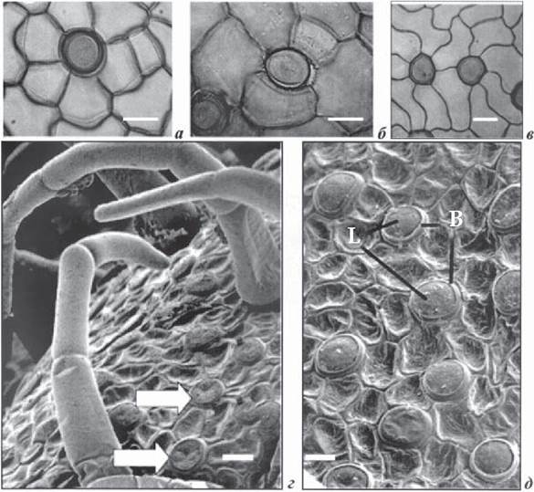
Fig. 1.7.3.1. Fragments of cells from the lower surface of hydrophyte leaves with hydropotes (a, b, c - light Microscopy after cuticle removal; d, e - scanning Electron microscopy: a - stephanocytic-type hydropote in Nuphar advena (Aiton) W.T. Aiton; b - hydropote complex with irregular architecture in N. polysepala; c - complex of two actinocytic hydropotes in N. caerulea Savigny; d - lower leaf surface of Victoria amazonica with numerous hydropotes lacking Hair-like apical parts (arrows) and several hydropotes with attached hair-like structures; e - basal part (B) and lens-shaped cell (L) of the V. amazonica hydropote complex (Carpenter, 2006). Scale bar = 20 µm
Trichomes also participate in regulating the efflux of apoplastic calcium onto the organ surface (DeSilva et al., 2001), thereby modulating stomatal function. Toxic components are likewise transported to the leaf surface via trichomes. Phenolic exudates from trichomes provide a first line of defense against microbial attack (Wagner et al., 2004). Researchers suggest that trichomes may also release oligosaccharins from the apoplast to protect leaves against infection. Recently, a novel diterpenoid (labdadiene diol) isolated from tobacco trichomes during viral infection was described. This diterpenoid activated protein kinase and enhanced resistance to viral infection. The authors suggest that labdadiene diol acts as a signaling molecule during pathogen interaction with The plant cell (Seo et al., 2003).
Hydropotes. Floating and submerged leaves, stems, and floral petals of heterophyllous aquatic plants are characterized by specialized structures known as hydropotes—a specific type of gland possessing dual secretory and absorptive Functions (Lüttge, Krapf, 1969; Wilkinson, 1979). Hydropotes are involved in ion and water transport over short distances (aquatic environment ↔ epidermal cells), intermediate distances (aquatic environment ↔ photosynthesizing leaf cells), and long distances (aquatic environment ↔ leaf ↔ petiole ↔ root) (Lüttge, Krapf, 1983). A hydropote is a structure located on the epidermal surface of leaves and stems, featuring unusual Cell wall labyrinths. Hydropotes are cup-shaped and project above the epidermis. Utilizing light and scanning electron microscopy to examine the lower and upper surfaces of floating leaves from six species—Nuphar advena, N. lutea, Nymphaea polysepala (Engelm.) Greene, Nymphaea caerulea, Nymphaea flava Aether ex Audubon, and Nymphaea nouchali Burm. f.—as well as the leaves of Victoria amazonica (Fig. 1.7.3.1), J. Carpenter (Carpenter, 2006) established that hydropotes consist of a single cell or a group of cells of the stephanocytic or actinocytic type, anchored by a subepidermal biconvex lens-shaped cell. Following the terminology of U. Lüttge et al. (Lüttge, Krapf, 1969; Wilkinson, 1979), this anchoring cell is termed the "FOOT" cell (Fig. 1.7.3.1), while the upper cell serves as the main cup-shaped cell of the hydropote complex, featuring a depression and thick cell walls. Additionally, some authors note that hydropotes can be found on both leaf surfaces across a wide range of aquatic angiosperms and water ferns (Kaul, 1976; Wilkinson, 1979; Carpenter, 2006). The size and density of hydropotes within the epidermis depend on the species and epidermal type. R. Kaul, in his study of hydathodes across heterophyllous and homophyllous species from various genera, suggested that the hydropote, as an epidermal structure, is a prime example of convergent evolution in aquatic plants (Kaul, 1976).
Last update: 07/08/2026
Editorial and Educational Adaptation: This material has been compiled based on the primary/original source text. The project team performed an editorial review, corrected technical inaccuracies, structured sections, and adapted the content for an educational format.
What was processed:
- elimination of formatting defects (OCR errors, structural breaks, corrupted characters);
- editorial organization of content;
- standardization of terminology in accordance with academic sources;
- verification of factual statements against the original source text.
All mentions of the author, publication year, and origin of the primary text have been preserved in accordance with the source.